 Back
BackSynaptic Transmission & Neural Integration: Study Guide for ANP College Students
Study Guide - Smart Notes
 Tailored notes based on your materials, expanded with key definitions, examples, and context.
Synaptic Transmission & Neural Integration
Overview of Synapses in the Nervous System
Synapses are specialized junctions through which neurons signal to each other and to non-neuronal cells such as muscles or glands. They are essential for neural communication and integration within the nervous system.
Electrical Synapses: Utilize gap junctions for direct electrical coupling between cells. These are less common and typically found in cells with similar functions. The gap junction distance is about 30-50 nm.
Chemical Synapses: Use neurotransmitters to communicate with neurons, muscles, or glands. These are the most common type of synapse.
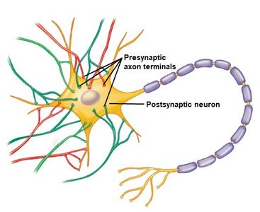
Presynaptic neuron: The neuron sending the signal.
Postsynaptic neuron: The neuron receiving the signal.
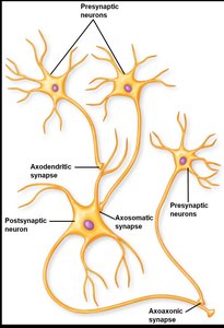
Functional Anatomy of Chemical Synapses
Chemical synapses involve the release of neurotransmitters from the presynaptic neuron, which bind to receptors on the postsynaptic neuron, leading to a response.
Axon terminal: Contains synaptic vesicles filled with neurotransmitters.
Voltage-gated Ca2+ channels: Open in response to an action potential, allowing Ca2+ influx.
Neurotransmitter release: Increased Ca2+ leads to more neurotransmitter exocytosis.
Reuptake molecules: Remove neurotransmitters from the synaptic cleft for recycling.
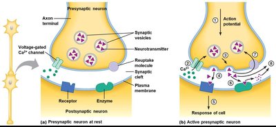
Clinical Correlation: SSRIs
Selective Serotonin Reuptake Inhibitors (SSRIs) are used to treat depression by preventing the reuptake of serotonin, thereby increasing its availability in the synaptic cleft.
Examples: Prozac, Lexapro, Paxil, Zoloft.
Mechanism: SSRIs block the reuptake of serotonin into the presynaptic neuron.
Signal Transduction at Chemical Synapses
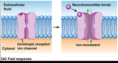
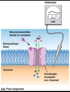
Fast Response: Ionotropic Receptors
Ionotropic receptors are ligand-gated ion channels that mediate rapid changes in membrane potential when neurotransmitters bind.
Mechanism: Neurotransmitter binds, ion channel opens, ions move across membrane.
Result: Change in membrane potential (Vm), called a post-synaptic potential (PSP).
PSP: Graded by the amount of neurotransmitter bound to receptors.
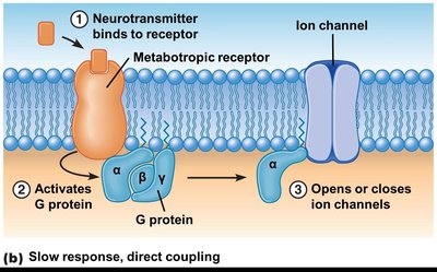
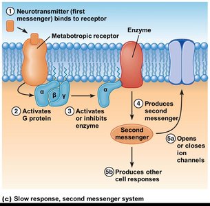
Slow Response: Metabotropic Receptors
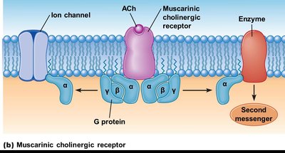
Metabotropic receptors are G protein-coupled receptors that mediate slower, longer-lasting changes in the postsynaptic cell.
Direct Coupling: G protein directly opens or closes ion channels.
Second Messenger System: G protein activates an enzyme, producing a second messenger (e.g., cAMP), which then affects ion channels or other cellular responses.
Excitatory and Inhibitory Synapses
Excitatory Synapses (EPSP)
Excitatory post-synaptic potentials (EPSPs) depolarize the postsynaptic membrane, bringing it closer to threshold for action potential generation.
Mechanism: Opening of Na+ or Ca2+ channels.
Result: Depolarization of Vm.
Inhibitory Synapses (IPSP)
Inhibitory post-synaptic potentials (IPSPs) hyperpolarize or stabilize the membrane potential, moving it further from threshold and reducing the likelihood of action potential generation.
Mechanism: Opening of K+ or Cl- channels.
Result: Hyperpolarization or stabilization of Vm.
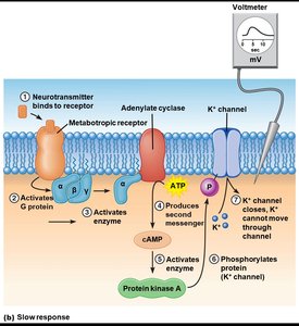
Slow Response Example: Closure of K+ Channels
Closure of K+ channels prevents cations from leaving the cell, resulting in depolarization.
Second messenger (cAMP): Produced by adenylate cyclase, activates protein kinase A, which phosphorylates K+ channels.
Neural Integration
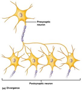
Divergence and Convergence
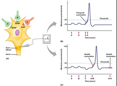
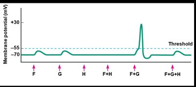
Neural integration involves the summation of inputs from multiple synapses to determine whether an action potential will be generated.
Divergence: One presynaptic neuron influences multiple postsynaptic neurons.
Convergence: Multiple presynaptic neurons influence a single postsynaptic neuron.
Summation: Temporal and Spatial
Summation is necessary for the postsynaptic neuron to reach threshold and trigger an action potential.
Temporal Summation: Multiple EPSPs from a single presynaptic neuron in rapid succession.
Spatial Summation: EPSPs from multiple presynaptic neurons occurring simultaneously.
Threshold: The membrane potential at which an action potential is triggered, typically around -55 mV.
Frequency Coding
Frequency coding refers to the relationship between the frequency of action potentials and the amount of neurotransmitter released.
More neurotransmitter released: Higher frequency of action potentials leads to more neurotransmitter exocytosis.
More voltage-gated Ca2+ channels open: Increased Ca2+ influx enhances neurotransmitter release.
Greater depolarization: Results in stronger postsynaptic responses.
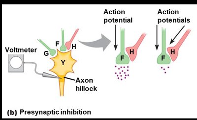
Presynaptic Modulation at Axoaxonic Synapses
Presynaptic Facilitation
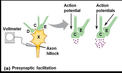
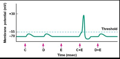
Presynaptic facilitation occurs when one neuron enhances the neurotransmitter release of another neuron at the axon terminal.
Mechanism: Increased Ca2+ influx and neurotransmitter release.
Presynaptic Inhibition
Presynaptic inhibition occurs when one neuron reduces the neurotransmitter release of another neuron at the axon terminal.
Mechanism: Decreased Ca2+ influx and neurotransmitter release.
Acetylcholine (ACh) and Cholinergic Receptors
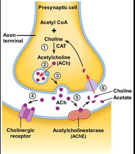
Synthesis and Breakdown of Acetylcholine
Acetylcholine is synthesized in the axon terminal cytosol and released from both CNS and PNS neurons. It is the most abundant neurotransmitter in the PNS.
Synthesis: Acetyl CoA + Choline (via choline acetyltransferase, CAT).
Breakdown: Acetylcholinesterase (AChE) breaks down ACh into choline and acetate.
Receptors: Binds to cholinergic receptors.
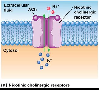
Cholinergic Receptors: Nicotinic and Muscarinic
Cholinergic receptors are classified as nicotinic or muscarinic, each with distinct mechanisms and locations.
Nicotinic receptors: Ionotropic, fast response, found in skeletal muscle and CNS.
Muscarinic receptors: Metabotropic, slow response, found in CNS and target organs of the PNS.
Key Concepts Table: Synaptic Transmission Types
Type | Mechanism | Speed | Example |
|---|---|---|---|
Ionotropic (Fast) | Ligand-gated ion channel | Milliseconds | Nicotinic ACh receptor |
Metabotropic (Slow, Direct) | G protein directly opens/closes channel | Seconds | Muscarinic ACh receptor |
Metabotropic (Slow, 2nd Messenger) | G protein activates enzyme, produces second messenger | Seconds to minutes | Muscarinic ACh receptor |
Additional info: The action of any chemical messenger depends on the type of receptor to which it binds, not the nature of the messenger itself.
