 Back
BackSynaptic Transmission: Electrical and Chemical Synapses in the Nervous System
Study Guide - Smart Notes
 Tailored notes based on your materials, expanded with key definitions, examples, and context.
CH 11 PT 4 - Synaptic Transmission in the Nervous System
Overview of Synapses
Synapses are specialized junctions through which neurons communicate with each other or with effector cells. There are two main types: electrical synapses and chemical synapses. Each type has distinct structures and mechanisms for transmitting signals.
Electrical Synapses
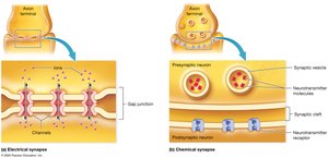
Electrical synapses allow direct, rapid transmission of electrical signals between neurons via gap junctions. These are less common than chemical synapses but are crucial for synchronizing activity in certain tissues.
Structure: Adjacent neurons are connected by gap junctions, which are protein channels that permit ions to flow directly from one cell to another.
Speed: Transmission is nearly instantaneous, with a delay of less than 0.1 ms.
Synchronization: Enables groups of cells to fire action potentials in unison, important for automatic behaviors (e.g., breathing), cardiac muscle, visceral smooth muscle, and developing nervous tissue.
Bidirectionality: Electrical signals can travel in both directions across the synapse.
Chemical Synapses
Chemical synapses are the predominant type in the nervous system. They convert electrical signals into chemical signals using neurotransmitters, allowing for more complex and modifiable communication.
Structure: Consist of a presynaptic axon terminal containing synaptic vesicles (filled with neurotransmitters), a synaptic cleft (20–50 nm wide), and a postsynaptic membrane with neurotransmitter receptors.
Unidirectionality: Signals are transmitted from the presynaptic to the postsynaptic neuron only.
Synaptic Delay: There is a brief delay (~0.5 ms) due to the time required for neurotransmitter release and binding.
Signal Modulation: Chemical synapses can vary in strength and effect, depending on the amount and type of neurotransmitter released and the receptors present.
Events at a Chemical Synapse
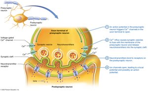
An action potential arrives at the presynaptic neuron's axon terminal, opening voltage-gated calcium ion channels.
Calcium influx triggers synaptic vesicles to fuse with the presynaptic membrane, releasing neurotransmitters into the synaptic cleft.
Neurotransmitters diffuse across the cleft and bind to receptors on the postsynaptic membrane (often ligand-gated ion channels).
Ion channels open, generating a local potential that may trigger an action potential in the postsynaptic neuron.
Postsynaptic Potentials
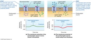
When neurotransmitters bind to receptors on the postsynaptic neuron, they cause local changes in membrane potential called postsynaptic potentials (PSPs). These can be excitatory or inhibitory:
Excitatory Postsynaptic Potential (EPSP): A small, local depolarization that brings the membrane potential closer to threshold, increasing the likelihood of an action potential. Typically caused by opening ligand-gated Na+ or Ca2+ channels.
Inhibitory Postsynaptic Potential (IPSP): A small, local hyperpolarization that moves the membrane potential further from threshold, decreasing the likelihood of an action potential. Caused by opening ligand-gated K+ channels (K+ efflux) or Cl− channels (Cl− influx).
Summation and Neural Integration
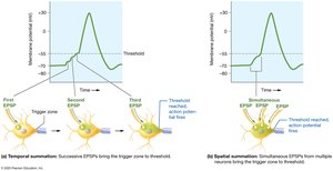
Neurons integrate multiple synaptic inputs through summation, which determines whether the postsynaptic neuron reaches threshold to fire an action potential. Summation can be temporal or spatial:
Temporal Summation: Rapid, successive EPSPs from a single presynaptic neuron add together if they occur within a short time frame (each EPSP lasts ~15 ms).
Spatial Summation: Simultaneous EPSPs from multiple presynaptic neurons combine at the postsynaptic membrane.
Both EPSPs and IPSPs can summate, and their combined effect determines the postsynaptic neuron's response.
Types of Channels and Pumps in Neuron Membranes
Neuronal function depends on the distribution and activity of various ion channels and pumps:
Ligand-Gated Ion Channels: Located on dendrites and cell bodies; open in response to neurotransmitter binding.
Voltage-Gated Na+ and K+ Channels: Found along the axon; responsible for generating and propagating action potentials.
Voltage-Gated Ca2+ Channels: Located at axon terminals; trigger neurotransmitter release via exocytosis.
Leak Channels and Na+/K+ Pump: Present throughout the neuron; maintain resting membrane potential and ion gradients.
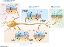
Table: Comparison of Electrical and Chemical Synapses
Feature | Electrical Synapse | Chemical Synapse |
|---|---|---|
Structure | Gap junctions | Synaptic cleft, vesicles, receptors |
Speed | Very fast (<0.1 ms) | Slower (~0.5 ms delay) |
Directionality | Bidirectional | Unidirectional |
Signal Modulation | Same size, less flexible | Variable size, diverse effects |
Location | Brainstem, heart, embryo | Most of nervous system |
Key Equations
Resting Membrane Potential: (Nernst equation for potassium)
Summation of Postsynaptic Potentials:
Additional info: The above equations are provided for academic context; the Nernst equation is simplified for potassium, and the summation equation illustrates the net effect of EPSPs and IPSPs on membrane potential.
