 Back
BackSynaptic Transmission: Structure, Function, and Mechanisms
Study Guide - Smart Notes
 Tailored notes based on your materials, expanded with key definitions, examples, and context.
Synaptic Transmission: Presynaptic Mechanisms
Overview of Synaptic Transmission
Synaptic transmission is the process by which neurons communicate with each other at specialized junctions called synapses. There are two main types of synapses:
Electrical synapses: Allow direct ionic current flow between neurons via gap junctions.
Chemical synapses: Involve the release of neurotransmitters from the presynaptic neuron to the postsynaptic neuron.
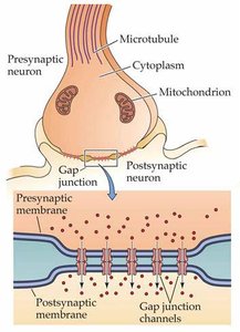
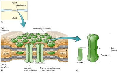
Structure and Function of Gap Junctions (Electrical Synapses)
Electrical synapses are formed by gap junctions, which are specialized channels that connect the cytoplasm of two adjacent neurons.
Connexon channels are embedded in both pre- and postsynaptic membranes.
The pore of a connexon channel is larger than typical ion channels, allowing passage of ions and larger molecules (e.g., ATP, second messengers).
Electrical synapses enable bi-directional transmission and are very rapid, with little to no delay.
They play a role in synchronized electrical activity, especially during embryonic development and in certain neural circuits.
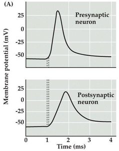
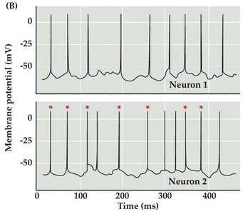
Electrophysiological Features of Gap Junctions
Simultaneous changes in membrane potential in coupled neurons.
Rapid transmission with minimal delay.
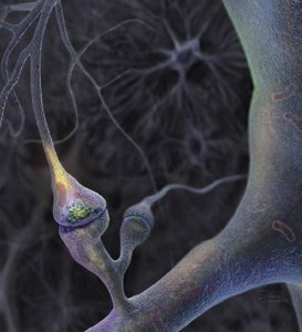
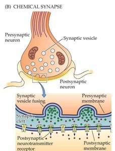
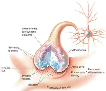
Structure and Function of Chemical Synapses
Chemical synapses involve the release of neurotransmitters from the presynaptic neuron, which bind to receptors on the postsynaptic neuron, resulting in a change in its properties.
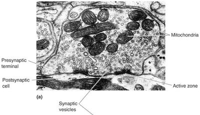
Presynaptic active zone: Site of neurotransmitter release.
Postsynaptic density (PSD): Site containing neurotransmitter receptors.
Synaptic vesicles: Store neurotransmitters.
Mitochondria: Provide ATP for metabolic support and regulate Ca2+.
Specificity and Features of Chemical Synaptic Transmission
Discrete molecular events are required for transmission.
Unidirectional signaling from presynaptic to postsynaptic cell.
Presynaptic calcium rise is critical for neurotransmission.
Postsynaptic responses can be excitatory or inhibitory, rapid or slow.
Synaptic transmission can be modulated and is subject to plasticity.
Quantal Release of Neurotransmitters
Historical Perspective: Katz's Experiments
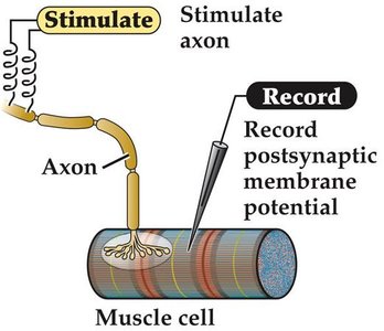
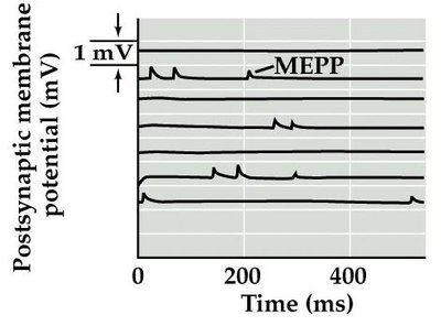
Bernard Katz's experiments at the frog neuromuscular junction (NMJ) demonstrated that neurotransmitter release occurs in discrete packets, or quanta.
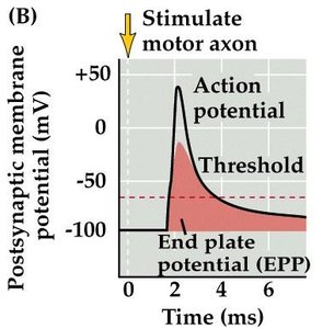
End-plate potentials (EPPs): Large, stimulus-evoked depolarizations in muscle fibers.
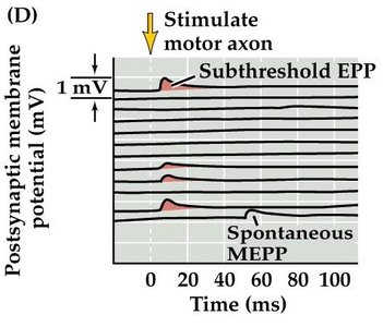
Miniature end-plate potentials (MEPPs): Small, spontaneous depolarizations observed without stimulation.
EPP amplitudes are multiples of MEPP amplitudes, suggesting quantal release.


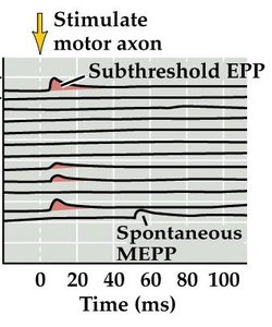
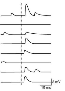
EPPs in Low Calcium Conditions
Lowering extracellular Ca2+ reduces release probability, resulting in subthreshold EPPs and more frequent MEPPs. 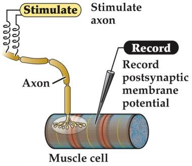
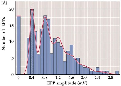
Quantal Analysis and Statistical Modeling
EPP amplitudes are not continuously variable but occur in specific multiples.
Each quantum corresponds to the release of a single synaptic vesicle.
Quantal release events are independent and follow a Poisson distribution.


Criteria for Defining a Neurotransmitter
A substance is considered a neurotransmitter if it meets the following criteria:
It is synthesized and present within the presynaptic terminal in sufficient amounts to produce a response.
It is released in response to presynaptic depolarization, and release is Ca2+-dependent.
Specific receptors for the substance are present on the postsynaptic cell.
Types of Neurotransmitters
Amino acid transmitters: Glutamate, GABA, Glycine, etc.
Monoamine transmitters: Dopamine, Norepinephrine, Serotonin, etc.
Polypeptide transmitters: Substance P, Endorphin, Neuropeptide Y, etc.
Other: Acetylcholine, Nitric oxide, ATP, etc.
Small-Molecule Neurotransmitters vs. Neuropeptides
Small-molecule transmitters are synthesized and packaged in the presynaptic terminal.
Neuropeptides are synthesized in the cell body, packaged into large dense-core vesicles, and transported to terminals.
Differential Release of Neurotransmitters
A single neuron can release both small-molecule neurotransmitters and neuropeptides, depending on activity patterns, Ca2+ concentration, and specific release machinery.
Synaptic Vesicle Cycle
The synaptic vesicle cycle describes the steps involved in neurotransmitter release and vesicle recycling:
Neurotransmitter is synthesized and stored in synaptic vesicles.
An action potential invades the presynaptic terminal.
Depolarization opens voltage-gated Ca2+ channels (VGCCs).
Ca2+ influx triggers vesicle fusion with the presynaptic membrane.
Neurotransmitter is released into the synaptic cleft via exocytosis.
Neurotransmitter binds to receptors on the postsynaptic membrane.
Current flows through neurotransmitter receptors, leading to postsynaptic depolarization.
Vesicle membrane is retrieved via endocytosis and recycled.
Molecular Machinery Underlying Neurotransmitter Release: The SNARE Complex
Docking: Vesicular SNARE (synaptobrevin) contacts plasma membrane SNAREs (syntaxin, SNAP-25).
Priming: SNARE proteins form a helical complex, bringing the vesicle close to the membrane.
Ca2+ entry: Synaptotagmin (Ca2+ sensor) binds Ca2+ and changes conformation.
Fusion: SNARE complex drives membrane fusion and neurotransmitter release.
Endocytosis: Vesicle membrane is retrieved via clathrin-mediated endocytosis.
Role of Ca2+ in Neurotransmitter Release
Ca2+ influx is essential for vesicle fusion and neurotransmitter release.
Experimental manipulation (e.g., Ca2+ chelators, direct injection) confirms its importance.
Summary Table: Comparison of Electrical and Chemical Synapses
Feature | Electrical Synapse | Chemical Synapse |
|---|---|---|
Structure | Gap junctions (connexons) | Synaptic cleft, vesicles, receptors |
Transmission | Direct ionic current | Neurotransmitter release |
Speed | Very rapid | Slower, with delay |
Directionality | Bi-directional | Unidirectional |
Plasticity | Limited | High (modulation, plasticity) |
Key Equations
Poisson Distribution for Quantal Release:
Where k is the number of quanta released, \lambda is the average number of quanta per stimulus.
Example: Neuromuscular Junction
Stimulation of a motor axon leads to neurotransmitter release at the NMJ, causing muscle contraction.
MEPPs and EPPs can be recorded to study quantal release.
Additional info:
Synaptic transmission is fundamental to nervous system function, underlying processes such as learning, memory, and coordinated movement.
Synaptic plasticity refers to activity-dependent changes in synaptic strength and structure.
