 Back
BackThe Anatomy and Physiology of Hearing and Equilibrium
Study Guide - Smart Notes
 Tailored notes based on your materials, expanded with key definitions, examples, and context.
The Special Senses: Hearing and Equilibrium
Overview of Hearing and Equilibrium
Hearing and equilibrium are two essential special senses provided by the internal ear. Both rely on mechanoreceptors known as hair cells, which detect sound waves and changes in head position. - Hearing: Enables detection and interpretation of sound waves. - Equilibrium: Provides information about the body's position in space, maintaining balance. - Hair cells: Specialized mechanoreceptors for both senses.
Anatomy of the Ear
The ear is divided into three anatomical regions, each with distinct functions and structures. - External ear: Collects sound waves and directs them inward. - Middle ear: Amplifies and transmits sound waves. - Internal ear: Contains sensory organs for hearing and equilibrium. 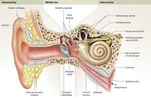
External Ear-collecting sound waves
The external ear is the visible portion and serves as the entry point for sound waves. - Auricle (pinna): Fleshy structure that collects and directs sound waves. - External acoustic meatus (auditory canal): Contains ceruminous glands that secrete cerumen (earwax), which helps to protect the ear); ends at the tympanic membrane. - Tympanic membrane (eardrum(just a membrane): Thin sheet that vibrates when struck by sound waves, separating the external and middle ear. 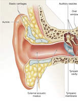
Middle Ear
The middle ear, or tympanic cavity, is an air-filled chamber that connects the external and internal ear. - Auditory tube (Eustachian tube): Connects to the pharynx, allowing pressure equalization across the eardrum. - Auditory ossicles: Three small bones (malleus, incus, stapes) that transmit and amplify sound waves. 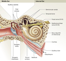
Auditory Ossicles
The ossicles are essential for transmitting sound vibrations from the tympanic membrane to the internal ear. - Malleus (hammer): Attaches to the eardrum. - Incus (anvil): Connects malleus to stapes. - Stapes (stirrup): Fills the oval window, transmitting vibrations to the internal ear. 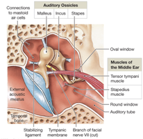
Internal Ear and Its Labyrinths
The internal ear contains the sensory organs for hearing and equilibrium, protected by the bony labyrinth. - Bony labyrinth: Fused with temporal bone, surrounds and protects the membranous labyrinth. - Membranous labyrinth: Tubes and chambers filled with endolymph; perilymph flows between the two labyrinths. 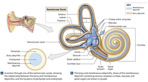
Three Parts of the Bony Labyrinth
1. Vestibule: Central region containing utricle and saccule; receptors for gravity and linear acceleration.
2. Semicircular canals(head moving itself: 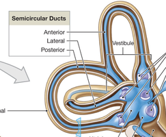
Three loop-shaped canals with semicircular ducts; detect rotational movements. 3. Cochlea(hearing): Spiral-shaped structure with cochlear duct and organ of Corti; converts sound vibrations into neural signals.
Inner Ear Receptors: Hair Cells
Hair cells are the primary sensory receptors of the inner ear, responsible for detecting mechanical(moving) changes. - Stereocilia: Hair-like projections on hair cells; bending alters neurotransmitter release. - Function: Direction of bending determines increase or decrease in neurotransmitter release, affecting signal transmission.
Equilibrium: Dynamic and Static
Equilibrium is maintained by hair cells in the vestibular complex, which includes the vestibule and semicircular canals. - Dynamic equilibrium: Maintains balance during motion; monitored by semicircular ducts. - Static equilibrium: Maintains balance and posture while motionless; monitored by saccule and utricle.
Semicircular Ducts
The three semicircular ducts are oriented in different planes, each responding to specific head movements. - Anterior duct: Nodding "yes". - Posterior duct: Tilting head side to side. - Lateral duct: Shaking head "no".
Semicircular Duct Anatomy
- Ampulla: Swollen base of each duct containing crista ampullaris. - Crista ampullaris: Raised structure with hair cells embedded in the cupula. - Function: Head rotation moves endolymph, pushing the cupula and bending stereocilia to activate hair cells.
Vestibule: Utricle and Saccule
- Utricle: Responds to gravity and horizontal acceleration; detects head tilt in the horizontal plane. - Saccule: Responds to gravity and vertical acceleration; detects head tilt in the vertical plane. - Maculae: Structures containing hair cells embedded in a gelatinous layer with otoliths (calcium carbonate crystals). - Function: Head movement shifts otoliths, bending stereocilia and altering nerve signals.
Pathways for Equilibrium Sensations
Sensory neurons from the vestibule and semicircular ducts form the vestibular nerve connecting inner ear to brain (branch of CN VIII). - Vestibular nuclei: Integrate balance information, send signals to cerebellum and cerebral cortex, and control eye, head, and neck movements.
Hearing: Detection of Sound
Hearing receptors are located in the cochlear duct. - Sound waves: Cause the tympanic membrane and ossicles to vibrate, creating pressure waves in cochlear fluid. - Pitch: Determined by location of activated hair cells. - Volume: Determined by number of hair cells activated.
Cochlear Duct
The cochlear duct is divided into three chambers: - Scala vestibuli: Filled with perilymph. - Scala media (cochlear duct): Filled with endolymph; contains organ of Corti. - Scala tympani: Filled with perilymph.
Organ of Corti(what allows us to hear)
The organ of Corti is the sensory organ for hearing, located on the basilar membrane. - Hair cells: Rest on basilar membrane; stereocilia contact tectorial membrane. - Function: Sound vibrations move basilar membrane, bending hair cells into tectorial membrane and activating sensory neurons.
Hearing Process
The process of hearing involves several steps: 1. Sound waves strike tympanic membrane. 2. Tympanic membrane vibrates auditory ossicles. 3. Vibration of stapes applies pressure to perilymph of scala vestibuli. 4. Pressure waves distort basilar membrane. 5. Movement of basilar membrane vibrates hair cells against tectorial membrane, altering neurotransmitter release. 6. Impulses travel to CNS via cochlear branch of vestibulocochlear nerve (CN VIII).
Auditory Pathway
- Cochlear nerve: Joins vestibular nerve to form vestibulocochlear nerve (CN VIII). - Signal transmission: Signals travel to medulla oblongata, through brainstem, then to thalamus, and finally to auditory cortex in temporal lobes.
Effects of Aging on Special Senses
Aging affects all special senses, including hearing and equilibrium. - Smell and taste: Decreased sensitivity due to fewer receptors and taste buds. - Vision: Lens loses elasticity (presbyopia), transparency (cataracts), loss of rods, and retinal damage (macular degeneration). - Equilibrium: Reduced vestibular function, increased dizziness, balance problems, and higher risk of falls. - Hearing: Hearing loss (presbycusis), reduced elasticity of tympanic membrane, high-frequency sounds affected first.
Summary of Ear Functions
Function | Process |
|---|---|
Equilibrium | Head movement → endolymph moves → hair cells bend → vestibular nerve → brain → balance maintained |
Hearing | Sound waves → tympanic membrane → ossicles → oval window → cochlea → hair cells → vestibulocochlear nerve → brain |
Additional info: Academic context and expanded explanations were added to clarify the structure and function of the ear, as well as the pathways for hearing and equilibrium.
