 Back
BackThe Appendicular Skeleton: Structure and Function
Study Guide - Smart Notes
 Tailored notes based on your materials, expanded with key definitions, examples, and context.
The Appendicular Skeleton
Overview and Introduction
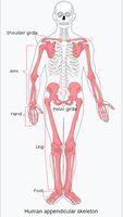
The appendicular skeleton comprises the bones that facilitate movement and manipulation of objects. It includes the bones of the limbs and the supporting girdles, accounting for approximately 60% of the bones in the human body.
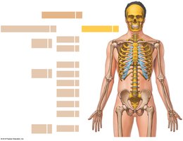
Key Point 1: The appendicular skeleton consists of the pectoral (shoulder) girdles, upper limbs, pelvic girdle, and lower limbs.
Key Point 2: It is essential for locomotion and interaction with the environment.
Example: The appendicular skeleton allows activities such as walking, grasping, and lifting.
Pectoral Girdle
Structure and Function
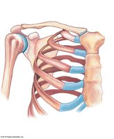
The pectoral girdle connects each arm to the body and positions the shoulder joint, providing a foundation for arm movement. Each girdle consists of one clavicle and one scapula, connecting to the axial skeleton only at the manubrium.
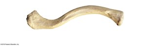
Clavicle: An S-shaped bone originating at the manubrium and articulating with the scapula at the acromial end.
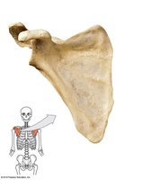
Scapula: A broad, flat triangle articulating with the humerus and clavicle. The anterior surface depression is the subscapular fossa.
Example: The shoulder joint (glenohumeral joint) is formed by the articulation of the scapula's glenoid cavity with the humerus.
Upper Limbs
Arm, Forearm, Wrist, and Hand
The upper limb skeleton includes the humerus (arm), ulna and radius (forearm), and the bones of the wrist and hand.
Humerus: The only bone in the arm, extending from the scapula to the elbow. Features include the head, greater and lesser tubercles, anatomical and surgical necks, deltoid tuberosity, and radial groove.
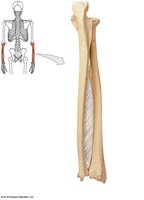
Ulna and Radius: The forearm consists of the medial ulna and lateral radius, connected by the interosseous membrane. The ulna features the olecranon, trochlear notch, and styloid process; the radius has a disc-shaped head, radial tuberosity, and styloid process.
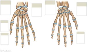
Carpal Bones: Eight carpal bones allow wrist movement, divided into proximal (scaphoid, lunate, triquetrum, pisiform) and distal (trapezium, trapezoid, capitate, hamate) groups.
Metacarpals and Phalanges: Five metacarpals form the palm, and phalanges make up the fingers. The thumb (pollex) has two phalanges; other fingers have three.
Pelvic Girdle
Structure and Function
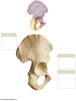
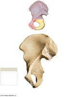
The pelvic girdle consists of two hip bones (coxal bones), each formed by the fusion of the ilium, ischium, and pubis. It attaches the lower limbs to the axial skeleton and supports body weight.
Acetabulum: The socket for the head of the femur, formed at the meeting point of the ilium, ischium, and pubis.
Obturator Foramen: A space encircled by the ischial and pubic rami, closed by collagen fibers for muscle attachment.
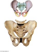
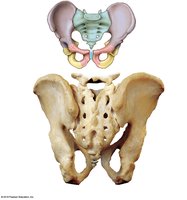
Pelvis: Includes the hip bones, sacrum, and coccyx, stabilized by ligaments and sacro-iliac joints.
Divisions of the Pelvis
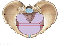
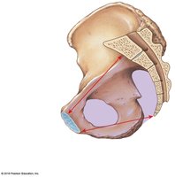
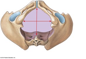
The pelvis is divided into the true pelvis (inferior to the pelvic brim, encloses the pelvic inlet) and the false pelvis (superior to the pelvic brim). The pelvic outlet is the opening bounded by the coccyx, ischial tuberosities, and pubic symphysis.
Perineum: The surface region bordered by the inferior edges of the pelvis, supported by perineal muscles.
Sex Differences in the Pelvis
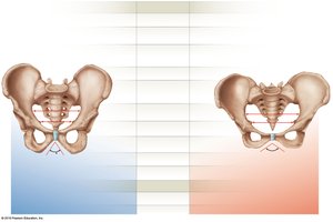
The female pelvis is broader, smoother, and lighter, with less prominent markings and a wider pelvic outlet compared to the male pelvis.
Comparison Table:
Feature | Male Pelvis | Female Pelvis |
|---|---|---|
General Appearance | Narrower, rougher, more robust | Broader, smoother, less robust |
Iliac Fossa | Deeper | Shallower |
Pelvic Inlet | Narrow, heart-shaped | Open, circular-shaped |
Pelvic Outlet | Narrow | Wide |
Pubic Angle | Less than 90º | 100º or more |
Lower Limbs
Thigh, Leg, Ankle, and Foot
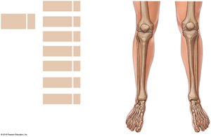
The lower limb skeleton includes the femur (thigh), patella (kneecap), tibia and fibula (leg), tarsal bones (ankle), metatarsals, and phalanges (foot).
Femur: The longest and heaviest bone, articulates with the hip at the acetabulum. Features include the head, neck, greater and lesser trochanters, linea aspera, epicondyles, and condyles.
Patella: A large sesamoid bone within the quadriceps tendon, articulates with the femur.
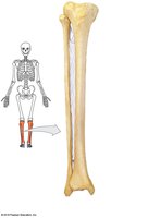
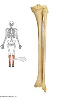
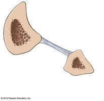
Tibia: The medial, weight-bearing bone of the leg, with medial and lateral condyles, tibial tuberosity, anterior margin, and medial malleolus.
Fibula: The lateral bone, with the head articulating with the tibia and lateral malleolus at the ankle.
Ankle and Foot
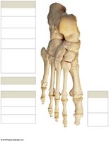
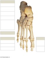
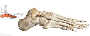
The ankle (tarsus) consists of seven tarsal bones, including the talus, calcaneus, cuboid, navicular, and three cuneiform bones. The foot has five metatarsals and 14 phalanges. The hallux (great toe) has two phalanges; other toes have three.
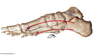
Arches of the Foot: The longitudinal arch ties the calcaneus to the distal metatarsals, while the transverse arch reflects the curvature from one side to the other.
Individual Skeletal Variation
Factors and Implications
Skeletal features can reveal muscle strength, medical history, sex, age, and body size. Modifications in the female pelvis for childbearing include the hormone relaxin, which loosens the pubic symphysis and sacro-iliac ligaments, increasing the size of the pelvic inlet and outlet.
Key Point: Skeletal variation is important for forensic and clinical assessments.
Example: The broader female pelvis facilitates childbirth.
Summary Table: Major Bones of the Appendicular Skeleton
Region | Main Bones |
|---|---|
Pectoral Girdle | Clavicle, Scapula |
Upper Limb | Humerus, Radius, Ulna, Carpals, Metacarpals, Phalanges |
Pelvic Girdle | Ilium, Ischium, Pubis |
Lower Limb | Femur, Patella, Tibia, Fibula, Tarsals, Metatarsals, Phalanges |
Additional info: The appendicular skeleton is essential for movement, manipulation, and support, and its structure reflects evolutionary adaptations for bipedal locomotion and dexterity.
