 Back
BackThe Appendicular Skeleton: Structure and Function
Study Guide - Smart Notes
 Tailored notes based on your materials, expanded with key definitions, examples, and context.
The Appendicular Skeleton
Overview and Primary Function
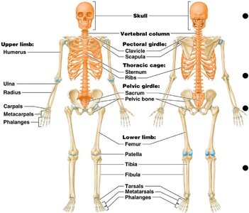
The appendicular skeleton is a major division of the human skeleton, primarily responsible for facilitating movement. It includes the bones of the upper and lower limbs, as well as the pectoral and pelvic girdles, which anchor the limbs to the axial skeleton.
Primary function: Aid in movement and locomotion.
Components:
Upper limbs (30 bones each)
Lower limbs (30 bones each)
Pectoral girdle (4 bones)
Pelvic girdle (2 bones)
Pectoral Girdle
Structure and Function
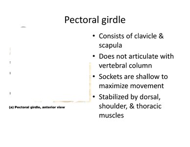
The pectoral girdle consists of the clavicle and scapula, providing attachment points for the upper limbs. It is designed for maximum mobility rather than stability, allowing a wide range of arm movements.
Composed of two bones: clavicle (collarbone) and scapula (shoulder blade).
Does not articulate with the vertebral column.
Sockets are shallow, maximizing movement but reducing stability.
Stabilized by dorsal, shoulder, and thoracic muscles.
Clavicle
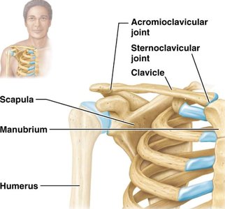
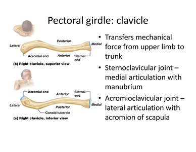
The clavicle acts as a strut to transfer mechanical force from the upper limb to the trunk. It forms two important joints:
Sternoclavicular joint: Medial articulation with the manubrium of the sternum.
Acromioclavicular joint: Lateral articulation with the acromion of the scapula.
Scapula
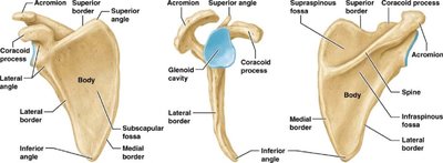
The scapula (shoulder blade) is a flat, triangular bone with several important surface features:
Spine: Large posterior process for muscle attachment.
Acromion: Flattened lateral portion of the spine, articulates with the clavicle.
Coracoid process: Anterior projection, inferior to the clavicle, for muscle attachment.
Glenoid cavity: Shallow socket that articulates with the head of the humerus to form the shoulder joint.
Upper Limb
Arm: Humerus
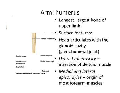
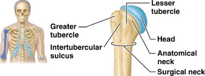
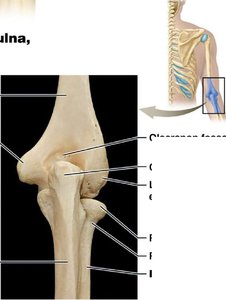
The humerus is the longest and largest bone of the upper limb. It articulates proximally with the scapula and distally with the radius and ulna.
Head: Articulates with the glenoid cavity (glenohumeral joint).
Deltoid tuberosity: Site of deltoid muscle insertion.
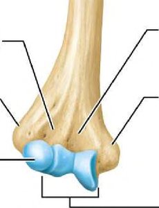
Medial and lateral epicondyles: Origins of most forearm muscles.
Forearm: Radius and Ulna
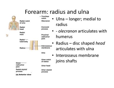
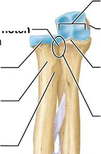
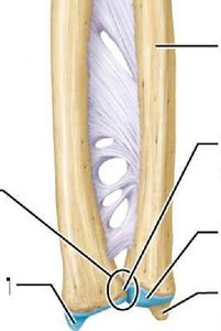
The ulna and radius are the two bones of the forearm. The ulna is longer and located medially, while the radius is shorter and lateral.
Ulna: Olecranon process articulates with the humerus.
Radius: Disc-shaped head articulates with the ulna and humerus.
Interosseous membrane: Connects the shafts of the radius and ulna.
Articulations at the Elbow
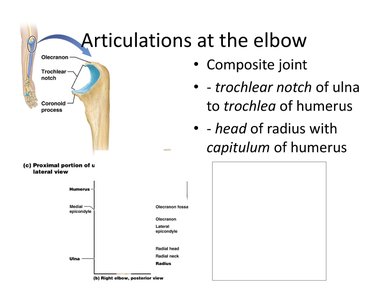
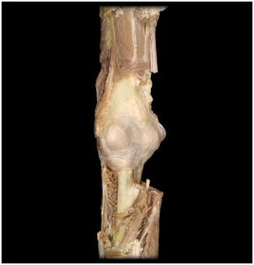
The elbow joint is a composite joint involving the humerus, radius, and ulna.
Trochlear notch of ulna articulates with the trochlea of humerus.
Head of radius articulates with the capitulum of humerus.
Hand: Carpals, Metacarpals, Phalanges
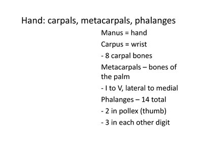
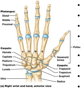
The hand consists of the carpus (wrist), metacarpals (palm), and phalanges (fingers).
Carpus: 8 carpal bones form the wrist.
Metacarpals: 5 bones numbered I to V, lateral to medial.
Phalanges: 14 bones; 2 in the thumb (pollex), 3 in each other digit.
Pelvic Girdle
Structure and Function
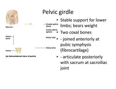
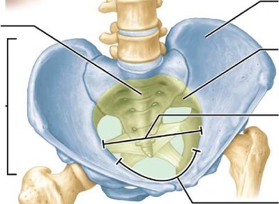
The pelvic girdle provides stable support for the lower limbs and bears the weight of the upper body. It consists of two coxal (hip) bones joined anteriorly at the pubic symphysis and articulating posteriorly with the sacrum at the sacroiliac joint.
Pubic symphysis: Fibrocartilage joint connecting the two coxal bones anteriorly.
Sacroiliac joint: Articulation with the sacrum posteriorly.
Os Coxa (Hip Bone)
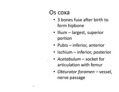
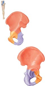
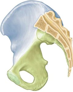
The os coxa forms by the fusion of three bones after birth:
Ilium: Largest, superior portion.
Pubis: Inferior, anterior portion.
Ischium: Inferior, posterior portion.
Acetabulum: Socket for articulation with the femur.
Obturator foramen: Passage for vessels and nerves.
Greater vs. Lesser Pelves
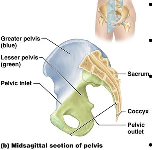
The pelvis is divided into the greater (false) pelvis and lesser (true) pelvis by the pelvic brim.
Pelvic brim: Ring from sacral promontory to pubic symphysis.
Lesser pelvis: Below brim; forms the birth canal.
Greater pelvis: Above brim; supports abdominal organs.
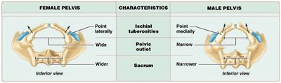
Female vs. Male Pelves
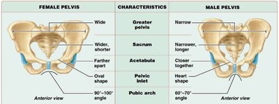
There are notable differences between female and male pelves, reflecting adaptations for childbirth in females.
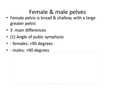
Angle of pubic symphysis: Females >90°, males <90°.
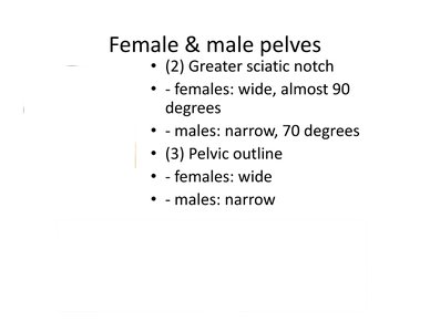
Greater sciatic notch: Females wide (~90°), males narrow (~70°).
Pelvic outline: Females wide, males narrow.
Feature | Female Pelvis | Male Pelvis |
|---|---|---|
Greater pelvis | Wide | Narrow |
Pelvic inlet | Oval | Heart-shaped |
Pubic arch | 90–100° | 60–70° |
Ischial tuberosities | Point laterally | Point medially |
Lower Limb
Thigh: Femur
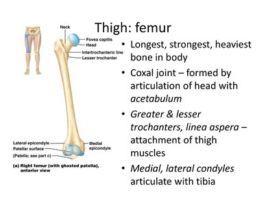
The femur is the longest, strongest, and heaviest bone in the body. It articulates proximally with the acetabulum of the hip bone and distally with the tibia.
Greater and lesser trochanters, linea aspera: Sites for muscle attachment.
Medial and lateral condyles: Articulate with the tibia.
Thigh, Kneecap: Patella
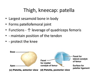
The patella is the largest sesamoid bone in the body, forming the patellofemoral joint. It increases the leverage of the quadriceps femoris muscle, maintains the position of the tendon, and protects the knee joint.
Leg: Tibia and Fibula
The tibia and fibula are the two bones of the lower leg. The tibia is larger and medial, bearing most of the body's weight, while the fibula is smaller and lateral.
Tibia: Articulates with the femur (at the knee) and talus (at the ankle).
Fibula: Bears little weight; lateral malleolus forms the outer part of the ankle.
Foot: Tarsals, Metatarsals, Phalanges
The foot consists of the tarsus (ankle), metatarsals (sole), and phalanges (toes).
Tarsus: 7 tarsal bones form the ankle.
Metatarsals: 5 bones numbered I to V, medial to lateral.
Phalanges: 14 bones; 2 in the hallux (big toe), 3 in each other digit.
Arches of the Foot
The foot has longitudinal (medial and lateral) and transverse arches, which support body weight, provide leverage, absorb shock, and flex during movement to redistribute body weight.
Longitudinal arch: Runs from heel to toe, divided into medial and lateral parts.
Transverse arch: Runs across the midfoot.
Example: The structure of the arches allows the foot to act as a spring during walking and running, enhancing efficiency and reducing injury risk.
