 Back
Ch 8:The Appendicular Skeleton: Structure and Function of the Limbs and Girdles
Study Guide - Smart Notes
 Tailored notes based on your materials, expanded with key definitions, examples, and context.
The Pectoral (Shoulder) Girdle
Structure and Function
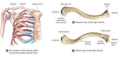
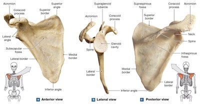
The pectoral girdle (shoulder girdle) attaches the upper limbs to the axial skeleton and provides mobility and flexibility to the shoulder region. It consists of two clavicles and two scapulae. The only direct bony connection to the axial skeleton is at the sternoclavicular joint, where the clavicle meets the manubrium of the sternum. The scapulae are supported and positioned by skeletal muscles, allowing for a wide range of motion but less strength compared to the pelvic girdle.
Clavicle: S-shaped bone connecting the sternum to the scapula; provides attachment for muscles and ligaments.
Scapula: Flat, triangular bone on the posterior thoracic cage; serves as a site for muscle attachment and articulates with the humerus at the glenoid cavity.
Mobility: The arrangement allows for extensive movement of the upper limb.
Bones of the Upper Limb
Humerus
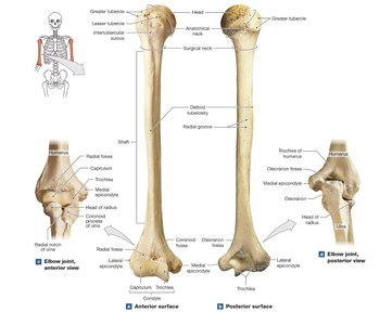
The humerus is the single bone of the arm, extending from the scapula to the elbow. It features a rounded head that articulates with the scapula, greater and lesser tubercles for muscle attachment, and a shaft with the deltoid tuberosity. The distal end forms the condyle, which articulates with the radius and ulna.
Greater and Lesser Tubercles: Sites for muscle attachment.
Deltoid Tuberosity: Attachment for the deltoid muscle.
Epicondyles: Medial and lateral projections for muscle attachment; the ulnar nerve passes behind the medial epicondyle ("funny bone").
Condyle: Divided into the trochlea (medial, articulates with ulna) and capitulum (lateral, articulates with radius).
Ulna and Radius
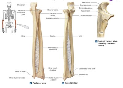
The ulna (medial) and radius (lateral) are parallel long bones of the forearm. The ulna forms the point of the elbow (olecranon) and articulates with the humerus at the trochlear notch. The radius articulates with the capitulum of the humerus and is important for wrist movement.
Ulna: Olecranon (elbow), trochlear notch, coronoid process, ulnar head, and styloid process.
Radius: Head, neck, radial tuberosity (biceps attachment), ulnar notch, and styloid process.
Interosseous Membrane: Connects the shafts of the radius and ulna.
Bones of the Wrist and Hand
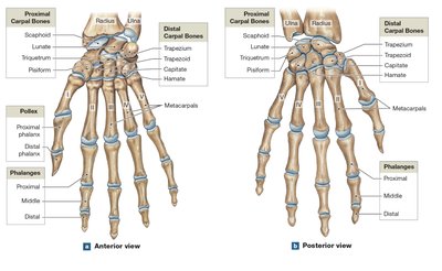
The wrist (carpus) contains eight carpal bones arranged in two rows. The hand consists of five metacarpals and fourteen phalanges (finger bones). The thumb (pollex) has two phalanges; other fingers have three each.
Proximal Carpals: Scaphoid, lunate, triquetrum, pisiform.
Distal Carpals: Trapezium, trapezoid, capitate, hamate.
Metacarpals: Numbered I–V from lateral (thumb) to medial.
Phalanges: Proximal, middle, and distal (except thumb).
The Pelvic Girdle
Structure and Function
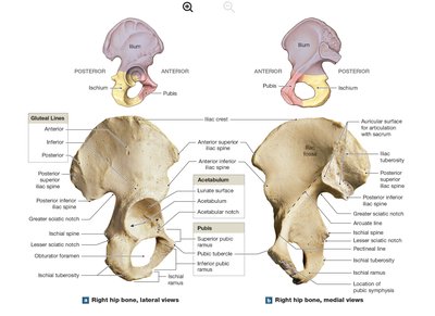
The pelvic girdle attaches the lower limbs to the axial skeleton and supports the weight of the upper body. It consists of two hip bones (coxal bones), each formed by the fusion of the ilium, ischium, and pubis. The pelvic girdle is more massive and less mobile than the pectoral girdle, reflecting its role in weight bearing and locomotion.
Acetabulum: Socket for articulation with the femoral head.
Ilium: Superior, broad portion; attachment for muscles and ligaments.
Ischium: Posterior, inferior part; supports body weight when sitting.
Pubis: Anterior, inferior part; forms the pubic symphysis with the opposite pubis.
Obturator Foramen: Large opening for nerves and blood vessels.
The Pelvis
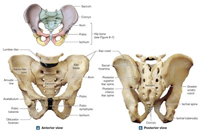
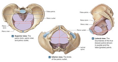
The pelvis is a composite structure including the pelvic girdle, sacrum, and coccyx. It is divided into the true (lesser) pelvis and false (greater) pelvis by the pelvic brim. The pelvic outlet is the inferior opening, important in childbirth. Ligaments and muscles stabilize the pelvis and support pelvic organs.
Sacroiliac Joint: Connects the ilium to the sacrum.
Pelvic Brim: Bony edge separating true and false pelvis.
Perineum: Region below the pelvic outlet, supporting pelvic organs.
Bones of the Lower Limb
Femur (Thighbone)
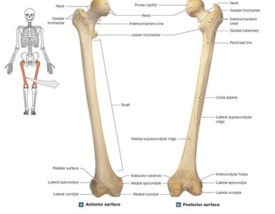
The femur is the longest and strongest bone in the body, articulating proximally with the hip at the acetabulum and distally with the tibia at the knee. Major features include the head, neck, greater and lesser trochanters, shaft, and condyles.
Head: Articulates with the acetabulum; fovea capitis is the ligament attachment site.
Trochanters: Muscle attachment sites.
Linea Aspera: Posterior ridge for muscle attachment.
Condyles: Medial and lateral, articulate with tibia.
Patella (Kneecap)
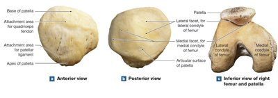
The patella is a sesamoid bone embedded in the quadriceps tendon. It protects the knee joint and improves the leverage of the quadriceps muscle. The posterior surface articulates with the femoral condyles.
Base: Superior attachment for quadriceps tendon.
Apex: Inferior attachment for patellar ligament.
Facets: Articulate with femoral condyles.
Tibia and Fibula (Leg)
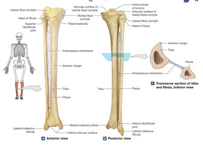
The tibia (medial) and fibula (lateral) support the leg. The tibia bears weight and articulates with the femur and ankle. The fibula provides muscle attachment and lateral stability to the ankle but does not bear significant weight.
Tibia: Medial and lateral condyles, tibial tuberosity (patellar ligament attachment), anterior margin, medial malleolus (ankle bump).
Fibula: Head, shaft, lateral malleolus (ankle stability).
Interosseous Membrane: Connects tibia and fibula.
Bones of the Ankle and Foot
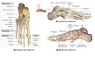
The ankle (tarsus) contains seven tarsal bones. The foot has five metatarsals and fourteen phalanges. The arrangement of bones forms arches that distribute body weight and provide shock absorption.
Tarsal Bones: Talus (articulates with tibia), calcaneus (heel), navicular, cuboid, and three cuneiforms (medial, intermediate, lateral).
Metatarsals: Numbered I–V from medial to lateral.
Phalanges: Hallux (great toe) has two; other toes have three each.
Arches: Longitudinal and transverse arches maintain foot shape and function.
Sex and Age Differences in the Skeleton
Sexual Dimorphism
Male and female skeletons differ in several ways, especially in the pelvis. Female pelves are generally wider, smoother, and have a larger pelvic inlet and outlet to facilitate childbirth. Other differences reflect variations in body size and muscle mass.
Pelvic Adaptations: Female pelvis adapted for childbirth; hormone relaxin increases pelvic flexibility during pregnancy.
Other Features: Skull, bone markings, and general size may differ between sexes.
Age-Related Changes
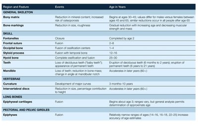
The skeleton undergoes predictable changes with age, including closure of growth plates (epiphyseal cartilages), reduction in bone mass, and fusion of certain bones. These changes can be used to estimate age at death in skeletal remains.
Region and Feature | Events | Age in Years |
|---|---|---|
General Skeleton: Bony matrix | Reduction in mineral content; increased risk of osteoporosis | Begins at age 30–45 |
Skull: Fontanelles | Closure | By age 2 |
Long Bones: Epiphyseal cartilages | Fusion | Begins about age 3; ends by late teens |
Pectoral and Pelvic Girdles: Epiphyses | Fusion | Ranges of ages (14–16, 18–21, 22–25) |
Vertebrae: Curvature | Development of major curves | 3 months–10 years |
Teeth | Eruption and loss of deciduous teeth; eruption of permanent teeth | 6 months–21 years (eruption); 60+ years (loss) |
Additional info: The timing of skeletal changes can vary due to genetics, nutrition, and health status. Heavy weight training in youth can damage growth plates, affecting adult stature. Degenerative changes such as osteoporosis are more common in older adults, especially postmenopausal women.
