 Back
The Appendicular Skeleton: Structure and Function
Study Guide - Smart Notes
 Tailored notes based on your materials, expanded with key definitions, examples, and context.
The Appendicular Skeleton
Overview
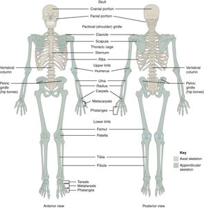
The appendicular skeleton is a major division of the human skeleton, consisting of 126 bones that form the limbs and the pectoral and pelvic girdles. It is essential for movement and interaction with the environment.
Definition: The appendicular skeleton includes the bones of the upper and lower limbs, as well as the girdles that attach them to the axial skeleton.
Components: Pectoral (shoulder) girdle, pelvic girdle, upper limbs, and lower limbs.
Function: Provides mobility, supports weight, and facilitates manipulation of objects.
The Pectoral Girdle
Structure and Articulations
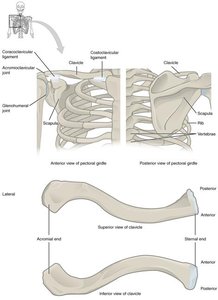
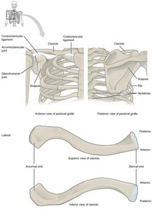
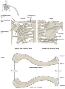
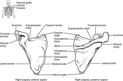
The pectoral girdle connects the upper limbs to the trunk and consists of the clavicles and scapulae.
Clavicle: S-shaped bone; medial end (sternal end) articulates with the manubrium of the sternum, forming the sternoclavicular joint (the only bony connection to the axial skeleton).
Lateral end (acromial end): Articulates with the acromion of the scapula, forming the acromioclavicular joint.
Scapula: Broad, flat, triangular bone; glenoid cavity articulates with the humerus to form the glenohumeral (shoulder) joint.
Bones of the Upper Limb
Regions and Major Bones
The upper limb is divided into three regions: arm (brachium), forearm (antebrachium), and hand. Each upper limb contains 30 bones.
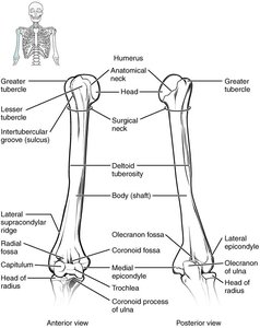
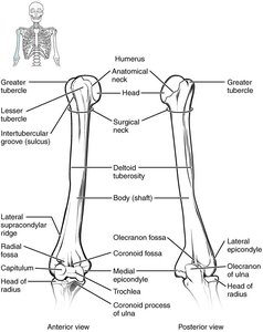
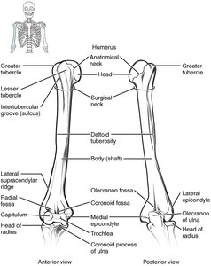
Arm: Contains the humerus, which articulates proximally with the scapula (glenoid cavity) and distally with the radius and ulna.
Forearm: Composed of the radius (lateral) and ulna (medial in anatomical position).
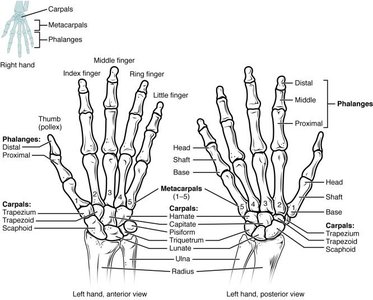
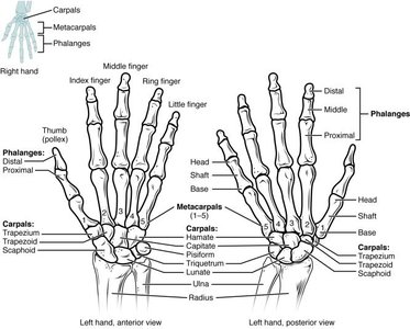
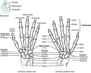
Hand: Includes carpal bones (wrist), metacarpals (palm), and phalanges (fingers and thumb).
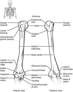
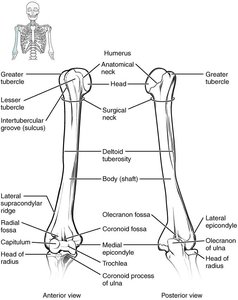
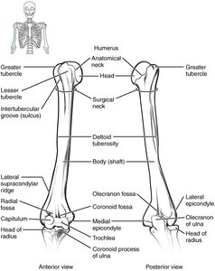
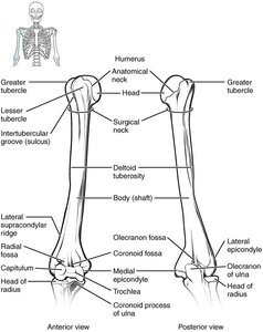
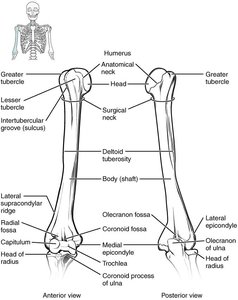
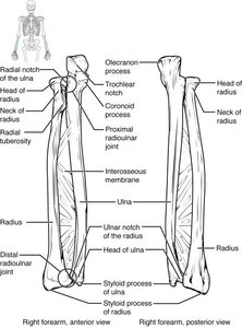
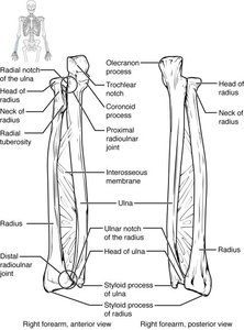
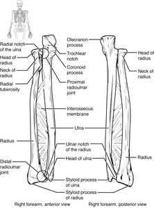
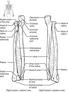
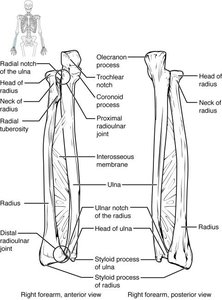
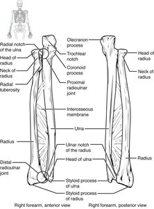
Forearm: Ulna and Radius
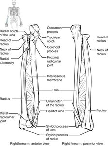
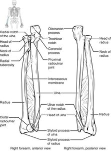
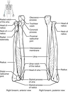
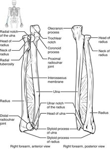
The ulna and radius are the two bones of the forearm, with the ulna being medial and the radius lateral in anatomical position.
Olecranon: Forms the point of the elbow; received by the olecranon fossa of the humerus when extended.
Trochlear notch: Articulates with the trochlea of the humerus.
Coronoid process: Received by the coronoid fossa of the humerus when flexed.
Radial notch: Site of articulation between the proximal radius and ulna (proximal radioulnar joint).
Styloid process: Attachment point for connective tissue.
Hand: Carpals, Metacarpals, and Phalanges
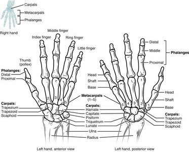
The hand consists of the carpus (wrist), metacarpals (palm), and phalanges (fingers and thumb).
Carpus: Eight carpal bones arranged in two rows (proximal: scaphoid, lunate, triquetrum, pisiform; distal: trapezium, trapezoid, capitate, hamate).
Metacarpals: Five bones, numbered 1–5 from the thumb.
Phalanges: Fourteen bones; thumb (pollex) has two (proximal and distal), fingers have three (proximal, middle, distal).
The Pelvic Girdle and Pelvis
Structure and Function
The pelvic girdle is composed of two hip (coxal) bones, each formed by the fusion of the ilium, ischium, and pubis.
Ilium: Superior and largest region; iliac crest is the superior margin.
Ischium: Posteroinferior region; ischial tuberosity bears weight when sitting.
Pubis: Anterior region; pubic symphysis unites left and right hip bones via fibrocartilage.
Acetabulum: Deep socket for articulation with the femur (hip joint).
Obturator foramen: Covered by collagen fibers; muscle attachment site.
Sacroiliac joint: Auricular surface of ilium articulates with sacrum, attaching pelvic girdle to axial skeleton.
Bones of the Lower Limb
Regions and Major Bones
The lower limb consists of 30 bones, including the femur, patella, tibia, fibula, tarsal bones, metatarsals, and phalanges.
Femur: Longest and heaviest bone; head articulates with acetabulum (hip joint).
Patella: Sesamoid bone; quadriceps tendon attaches superiorly, patellar ligament attaches inferiorly.
Tibia: Larger, medial bone; medial and lateral condyles articulate with femur (knee joint); tibial tuberosity is attachment site for patellar ligament; medial malleolus forms part of ankle joint.
Fibula: Slender, lateral bone; muscle attachment site; head articulates with tibia (proximal tibiofibular joint); lateral malleolus forms part of ankle joint.
Tarsus: Seven tarsal bones; talus articulates with tibia and fibula; calcaneus is the heel bone.
Metatarsals: Five bones, numbered 1–5 from medial side.
Phalanges: Fourteen bones; hallux (great toe) has two, other toes have three each.
Summary Table: Bones of the Appendicular Skeleton
Region | Main Bones | Key Joints |
|---|---|---|
Pectoral Girdle | Clavicle, Scapula | Sternoclavicular, Acromioclavicular, Glenohumeral |
Upper Limb | Humerus, Radius, Ulna, Carpals, Metacarpals, Phalanges | Shoulder, Elbow, Wrist, Radioulnar |
Pelvic Girdle | Ilium, Ischium, Pubis | Sacroiliac, Coxal (hip), Pubic Symphysis |
Lower Limb | Femur, Patella, Tibia, Fibula, Tarsals, Metatarsals, Phalanges | Hip, Knee, Ankle |
Key Terms and Definitions
Articulation: The point where two bones meet, forming a joint.
Girdle: A structure that supports and attaches limbs to the axial skeleton.
Sesamoid bone: A bone embedded within a tendon (e.g., patella).
Phalanx: Singular form of phalanges, the bones of fingers and toes.
Example: Functional Importance
The appendicular skeleton enables walking, running, grasping, and manipulation of objects, making it essential for daily activities and complex movements.
Additional info:
Some anatomical terms and joint names were expanded for clarity and completeness.
