 Back
The Appendicular Skeleton: Structure and Function of the Upper and Lower Limbs
Study Guide - Smart Notes
 Tailored notes based on your materials, expanded with key definitions, examples, and context.
Pectoral Girdle
Structure and Articulations
The pectoral girdle, also known as the shoulder girdle, is an incomplete ring of bones that connects the upper limb to the axial skeleton. It consists of the clavicle and scapula, and forms three major joints: the sternoclavicular, acromioclavicular, and glenohumeral joints. - Sternoclavicular joint: Medial end of clavicle articulates with the sternum. - Acromioclavicular joint: Lateral end of clavicle articulates with the scapula. - Glenohumeral joint: Scapula articulates with the humerus. 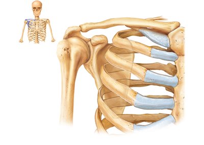
Clavicle
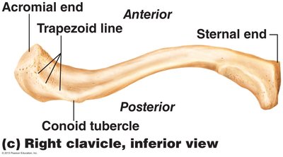
The clavicle, or collarbone, is an S-shaped bone that is easily palpated and acts as a brace for the shoulder. It is the most commonly fractured bone in the body. - Sternal (medial) end: Articulates with the sternum. - Acromial (lateral) end: Articulates with the scapula. - Conoid tubercle: Attachment site for ligaments. - Trapezoid line: Attachment site for ligaments. 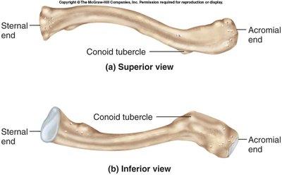
Scapula
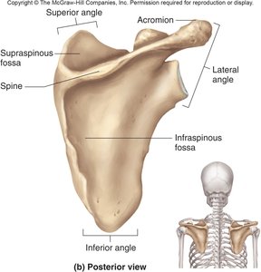
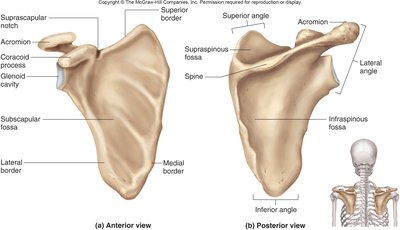
The scapula, or shoulder blade, is a flat, triangular bone that overlies ribs 2–7. It has several important anatomical landmarks: - Borders: Superior, medial, and lateral. - Angles: Superior, inferior, and lateral. - Coracoid process: Attachment for biceps brachii. - Glenoid cavity: Socket for the humerus. - Spine: Ridge on posterior surface. - Acromion: Apex of the shoulder. - Fossae: Supraspinous, infraspinous, and subscapular. 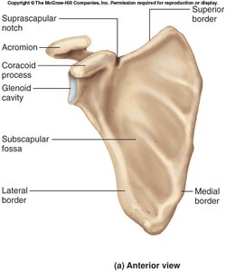
Upper Limb
Regions and Bones
The upper limb is divided into three regions and contains 30 bones per limb: - Brachium: Shoulder to elbow (humerus). - Antebrachium: Forearm (radius and ulna). - Manus: Hand (carpals, metacarpals, phalanges).
Humerus
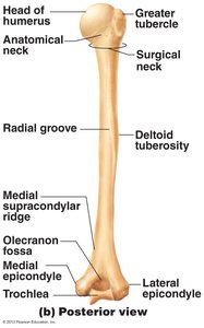
The humerus is the bone of the upper arm. Its proximal end articulates with the scapula, and its distal end forms the elbow joint. - Head: Articulates with glenoid cavity. - Intertubercular sulcus: For biceps tendon. - Capitulum: Articulates with radius. - Trochlea: Articulates with ulna. - Olecranon fossa: Deep posterior pit for olecranon process of ulna. 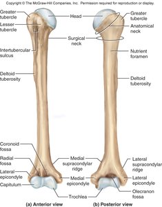
Radius and Ulna
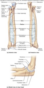
The radius and ulna are the bones of the forearm. They articulate with each other at the proximal and distal radioulnar joints. - Radius: Disc-shaped head for capitulum of humerus; styloid process at distal end. - Ulna: Olecranon and trochlear notch for humerus; radial notch for head of radius; styloid process at distal end. - Interosseous membrane: Connects the shafts of radius and ulna. 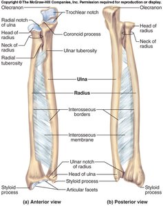
Hand: Carpals, Metacarpals, and Phalanges
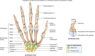
The hand consists of 27 bones: - Carpals: Eight bones arranged in two rows (proximal: scaphoid, lunate, triquetrum, pisiform; distal: trapezium, trapezoid, capitate, hamate). - Metacarpals: Five bones forming the palm, numbered I–V from thumb to little finger. - Phalanges: Bones of the fingers; thumb (pollex) has two, others have three (proximal, middle, distal). 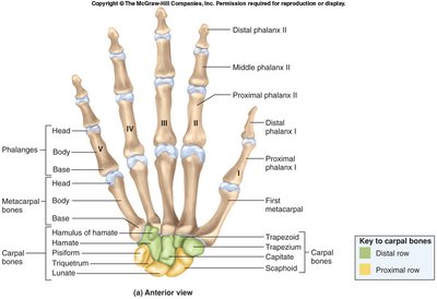
Pelvic Girdle
Structure and Joints
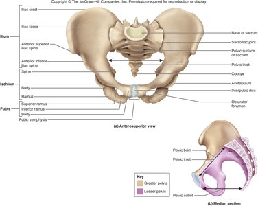
The pelvic girdle consists of two hip (coxal) bones and the sacrum. It forms the sacroiliac joints and the pubic symphysis. - Sacroiliac joint: Articulation between ilium and sacrum. - Pubic symphysis: Cartilaginous joint between pubic bones. 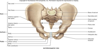
Hip Bone (Os Coxae)
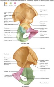
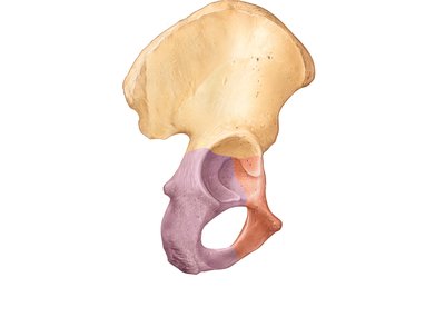
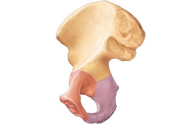
The hip bone is composed of three fused bones: ilium, ischium, and pubis. - Ilium: Iliac crest, iliac spines. - Ischium: Ischial tuberosity, ischial ramus. - Pubis: Pubic rami, pubic crest. - Acetabulum: Hip socket for femur. - Obturator foramen: Large opening for nerves and blood vessels. 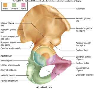
Pelvis Sexual Dimorphism
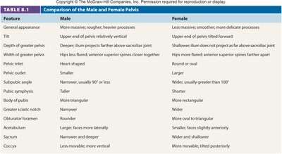
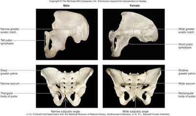
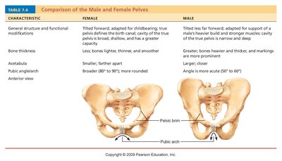
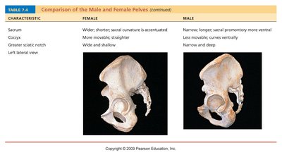
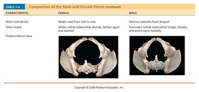
The pelvis exhibits sexual dimorphism, with differences between male and female pelves related to childbirth. - Female pelvis: Wider, shallower, larger pelvic inlet and outlet, adapted for childbirth. - Male pelvis: More robust, narrower, deeper, smaller pelvic inlet and outlet, adapted for support and muscle attachment. 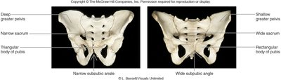
Lower Limb
Regions and Bones
The lower limb is divided into four regions and contains 30 bones per limb: - Femoral region: Thigh (femur, patella). - Crural region: Knee to ankle (tibia, fibula). - Tarsal region: Ankle (tarsal bones). - Pedal region: Foot (metatarsals, phalanges).
Femur and Patella
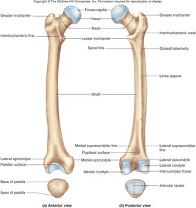
The femur is the longest and strongest bone in the body. The patella is the kneecap. - Femur: Head articulates with acetabulum; trochanters for muscle attachment; condyles and epicondyles at distal end. - Patella: Triangular sesamoid bone; apex is inferior. 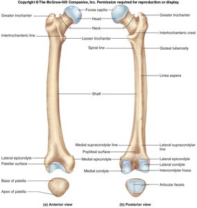
Tibia and Fibula
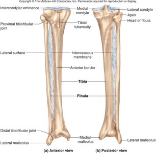
The tibia and fibula are the bones of the leg. - Tibia: Medial, weight-bearing bone; condyles, tuberosity, medial malleolus. - Fibula: Lateral, slender bone; head, lateral malleolus. - Interosseous membrane: Connects tibia and fibula. 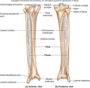
Ankle and Foot
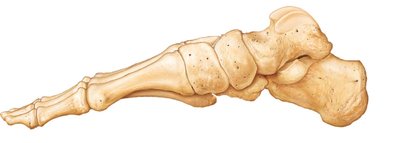
The ankle and foot are composed of tarsal bones, metatarsals, and phalanges. - Tarsals: Seven bones including calcaneus (heel), talus (most superior), navicular, cuboid, and three cuneiforms. - Metatarsals: Five bones, numbered I–V from medial to lateral. - Phalanges: Bones of the toes; hallux (great toe) has two, others have three (proximal, middle, distal). 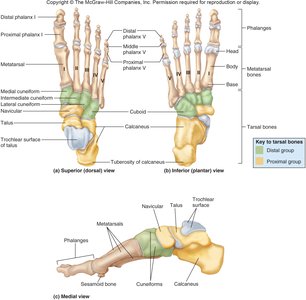
Developmental Aspects: Old Age
Changes in the Skeleton
With aging, several changes occur in the skeletal system: - Intervertebral discs: Become thin, less hydrated, and less elastic, increasing risk of herniation. - Loss of stature: Common by age 55 due to disc changes. - Costal cartilages: Ossify, making the thorax more rigid. - Bone mass: Decreases in all bones. Additional info: These changes contribute to decreased mobility and increased risk of fractures in the elderly.
