 Back
BackThe Appendicular Skeleton: Structure and Function
Study Guide - Smart Notes
 Tailored notes based on your materials, expanded with key definitions, examples, and context.
The Appendicular Skeleton
Overview of the Appendicular Skeleton
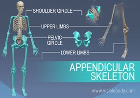
The appendicular skeleton consists of the bones of the limbs and the girdles that attach them to the axial skeleton. It is essential for movement and manipulation of the environment, as well as supporting the weight of the body during locomotion.
Pectoral (Shoulder) Girdle: Composed of the clavicle and scapula, connecting the upper limbs to the trunk.
Upper Limbs: Includes the humerus, radius, ulna, carpals, metacarpals, and phalanges.
Pelvic Girdle: Formed by the two hip bones (coxal bones), connecting the lower limbs to the trunk.
Lower Limbs: Includes the femur, tibia, fibula, tarsals, metatarsals, and phalanges.
Pectoral Girdle
Structure and Function
The pectoral girdle consists of two pairs of bones: the clavicles and scapulae. These bones form a nearly complete circle around the upper trunk and provide attachment points for muscles that move the upper limbs.
Clavicle (Collarbone): S-shaped bone that articulates medially with the sternum and laterally with the scapula. It acts as a brace to hold the arms laterally and serves as an attachment site for muscles.
Scapula (Shoulder Blade): Thin, triangular bone located on the dorsal side of the rib cage between ribs 2 and 7. Key features include the spine, acromion, coracoid process, and glenoid cavity (shoulder joint socket).
Upper Limb
Bones and Principal Markings
The upper limb is composed of 30 bones, divided into the arm, forearm, and hand.
Arm: The humerus is the only bone of the arm, articulating with the scapula at the shoulder and with the radius and ulna at the elbow.
Forearm: Consists of the radius and ulna, which are parallel and connected by the interosseous membrane. The ulna is primarily involved in forming the elbow joint, while the radius is involved in the wrist joint.
Hand: Contains 27 bones: 8 carpals (wrist), 5 metacarpals (palm), and 14 phalanges (fingers). The carpal bones are arranged in two rows, and the phalanges are numbered I-V from thumb to little finger.
Pelvic Girdle
Structure and Function
The pelvic girdle is formed by two hip bones (coxal bones), which unite anteriorly and with the sacrum posteriorly. It supports the weight of the upper body, attaches the lower limbs, and protects pelvic organs.
Ilium: The largest part, with the iliac crest and spines for muscle attachment.
Ischium: The postero-inferior part, featuring the ischial tuberosity (important for sitting).
Pubis: The anterior part, forming the pubic symphysis where the two pubic bones meet.
Acetabulum: The socket for the femur, formed by the fusion of the ilium, ischium, and pubis.
Pelvic Structure and Childbearing: The female pelvis is wider, shallower, and adapted for childbirth. The pelvic brim separates the false (upper) and true (lower) pelvis, with the true pelvis forming the birth canal.
Lower Limb
Bones and Principal Markings
The lower limb supports the body's weight and is adapted for locomotion. It consists of the thigh, leg, and foot.
Thigh: The femur is the largest and strongest bone, articulating with the acetabulum proximally and the tibia and patella distally.
Leg: Composed of the tibia (weight-bearing) and fibula (non-weight-bearing), connected by the interosseous membrane.
Foot: Contains 26 bones: 7 tarsals (including the calcaneus and talus), 5 metatarsals, and 14 phalanges. The foot has three arches (medial longitudinal, lateral longitudinal, and transverse) that help bear weight and provide flexibility.
Comparative Table: Upper vs. Lower Limb Bones
Region | Main Bones | Key Functions |
|---|---|---|
Upper Limb | Humerus, Radius, Ulna, Carpals, Metacarpals, Phalanges | Manipulation, grasping, fine motor skills |
Lower Limb | Femur, Tibia, Fibula, Tarsals, Metatarsals, Phalanges | Support, locomotion, weight-bearing |
Key Terms and Definitions
Glenoid Cavity: Shallow socket in the scapula for the head of the humerus.
Acetabulum: Deep socket in the hip bone for the head of the femur.
Interosseous Membrane: Fibrous sheet connecting parallel bones (e.g., radius and ulna; tibia and fibula).
Pelvic Brim: The edge separating the false and true pelvis.
Ischial Tuberosity: The part of the ischium that bears weight when sitting.
Summary
The appendicular skeleton is crucial for movement, support, and interaction with the environment. Understanding the structure and function of its components is essential for comprehending human anatomy and physiology, especially in relation to movement and weight-bearing activities.
