Back
BackThe Appendicular Skeleton: Structure and Function
Study Guide - Smart Notes
 Tailored notes based on your materials, expanded with key definitions, examples, and context.
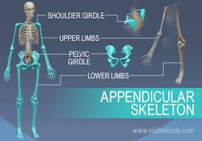
The Appendicular Skeleton
Overview of the Appendicular Skeleton
The appendicular skeleton consists of the bones of the limbs and the girdles that attach them to the axial skeleton. It is essential for movement and interaction with the environment, providing support and flexibility.
Pectoral (Shoulder) Girdle: Composed of the clavicle and scapula, connecting the upper limbs to the trunk.
Upper Limbs: Includes the humerus, radius, ulna, carpals, metacarpals, and phalanges.
Pelvic Girdle: Formed by the two hip bones (coxal bones), connecting the lower limbs to the trunk.
Lower Limbs: Includes the femur, tibia, fibula, tarsals, metatarsals, and phalanges.
Pectoral Girdle
Structure and Function
The pectoral girdle consists of two pairs of bones: the clavicles and scapulae. These bones form a nearly complete circle around the upper trunk and provide attachment points for muscles that move the upper limbs.
Clavicle (Collarbone): S-shaped bone that articulates medially with the sternum and laterally with the scapula. It acts as a brace to hold the arms laterally and provides muscle attachment points.
Scapula (Shoulder Blade): Thin, triangular bone located between ribs 2 and 7. It has three borders (superior, medial, lateral) and several important features, including the spine, acromion, coracoid process, and glenoid cavity (shoulder joint socket).
Upper Limb
Bones and Principal Markings
The upper limb is composed of 30 bones, divided into the arm, forearm, and hand.
Arm: The humerus is the only bone of the arm, articulating with the scapula, radius, and ulna. Key features include the head, anatomical and surgical necks, greater and lesser tubercles, deltoid tuberosity, and condyles (trochlea and capitulum).
Forearm: Consists of the radius and ulna, which are parallel long bones. The ulna is slightly longer and forms the elbow joint with the humerus, while the radius is involved in wrist articulation. The interosseous membrane connects the two bones.
Hand: Contains 27 bones: 8 carpals (wrist), 5 metacarpals (palm), and 14 phalanges (fingers). The carpal bones are arranged in two rows, and the phalanges are numbered I-V from thumb to little finger.
Pelvic Girdle
Structure and Function
The pelvic girdle is formed by two hip bones (coxal bones), which unite anteriorly and with the sacrum posteriorly. It supports the weight of the upper body, attaches the lower limbs, and protects pelvic organs.
Each coxal bone is formed by the fusion of three bones: ilium, ischium, and pubis.
The acetabulum is the socket where all three bones meet, forming the hip joint.
The pelvic brim separates the false pelvis (superior) from the true pelvis (inferior), which defines the birth canal.
Male vs. Female Pelvis
Female pelvis: Wider, shallower, lighter, and rounder, adapted for childbearing.
Male pelvis: Narrower and deeper, with a more pronounced pelvic inlet and outlet.
Lower Limb
Bones and Principal Markings
The lower limb supports the entire weight of the body and is adapted for locomotion. It consists of the thigh, leg, and foot.
Thigh: The femur is the largest and strongest bone in the body, articulating with the acetabulum proximally and the tibia and patella distally. Key features include the head, neck, trochanters, condyles, and epicondyles.
Leg: Composed of the tibia (weight-bearing) and fibula (non-weight-bearing), connected by the interosseous membrane. The tibia articulates with the femur and foot, while the fibula provides lateral stability.
Foot: Contains 26 bones: 7 tarsals (ankle), 5 metatarsals, and 14 phalanges (toes). The calcaneus (heel bone) and talus bear most of the body's weight. The foot has three arches (lateral longitudinal, medial longitudinal, and transverse) that help distribute weight and provide flexibility.
Summary Table: Major Bones of the Appendicular Skeleton
Region | Main Bones | Key Features |
|---|---|---|
Pectoral Girdle | Clavicle, Scapula | Glenoid cavity, acromion, coracoid process |
Upper Limb | Humerus, Radius, Ulna, Carpals, Metacarpals, Phalanges | Olecranon, trochlea, capitulum, interosseous membrane |
Pelvic Girdle | Ilium, Ischium, Pubis | Acetabulum, pelvic brim, pubic symphysis |
Lower Limb | Femur, Tibia, Fibula, Tarsals, Metatarsals, Phalanges | Greater/lesser trochanter, condyles, malleolus, arches |
Key Definitions
Glenoid cavity: Shallow socket in the scapula that articulates with the head of the humerus.
Acetabulum: Deep socket in the hip bone that receives the head of the femur.
Interosseous membrane: Fibrous sheet connecting the radius and ulna or tibia and fibula.
Pelvic brim: Bony rim separating the false and true pelvis.
Example: Clinical Relevance
Clavicle fractures are common due to its superficial position and function as a brace. The S-shape helps prevent damage to underlying vessels.
Pelvic differences are important in obstetrics for assessing childbirth risk.