 Back
The Appendicular Skeleton: Structure and Function
Study Guide - Smart Notes
 Tailored notes based on your materials, expanded with key definitions, examples, and context.
The Appendicular Skeleton
Overview
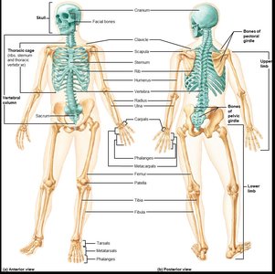
The appendicular skeleton consists of the bones of the limbs and their girdles, which are essential for movement, manipulation, and support of the body. It is divided into the pectoral girdle (upper limb attachment), upper limb, pelvic girdle (lower limb attachment), and lower limb.
Pectoral girdle: Attaches upper limbs to the body trunk.
Upper limb: Provides a wide range of motion and manipulation.
Pelvic girdle: Attaches lower limbs to the body trunk.
Lower limb: Built to bear the body’s weight and keep the body upright; important for locomotion.
Pectoral Girdle
Structure and Function
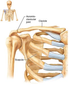
The pectoral girdle, or shoulder girdle, consists of the clavicles (anteriorly) and scapulae (posteriorly). It attaches the upper limbs to the axial skeleton and provides attachment sites for muscles that move the upper limbs.
Mobility: Scapulae are not attached to the axial skeleton, and the socket of the shoulder joint is shallow, allowing for a great degree of movement.
Clavicles
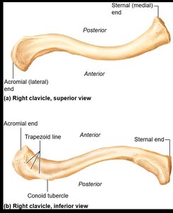
The clavicles, or collarbones, are S-shaped bones that anchor muscles and act as braces to hold the scapulae and arms out laterally.
Sternal end: Articulates with the sternum medially.
Acromial end: Articulates laterally with the scapula.
Scapulae
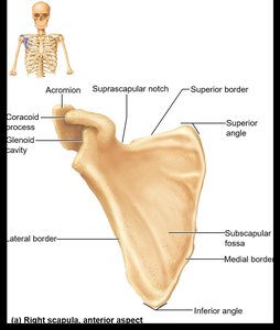
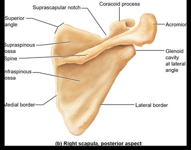
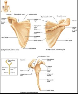
The scapulae, or shoulder blades, are thin, triangular bones located on the dorsal surface of the rib cage. Each scapula has three borders (superior, medial, lateral) and three angles (superior, lateral, inferior).
Spine: Prominent ridge on the posterior side.
Acromion: Lateral projection that forms the acromioclavicular joint.
Coracoid process: Anterior projection anchoring the biceps muscle.
Suprascapular notch: Opening for nerves.
Fossae: Large depressions for muscle attachment.
The Upper Limb
Bone Structure
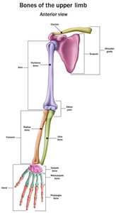
Each upper limb contains 30 bones, forming the arm, forearm, and hand.
Arm: Humerus
Forearm: Radius and ulna
Hand: 8 carpal bones (wrist), 5 metacarpal bones (palm), 14 phalanges (fingers)
Humerus
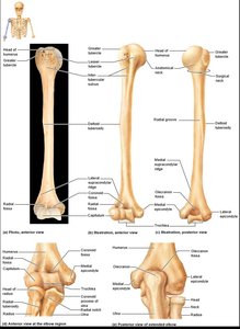
The humerus is the only bone of the arm and is the largest and longest bone of the upper limb.
Head: Fits into the glenoid cavity of the scapula.
Anatomical neck: Slight constriction below the head.
Greater and lesser tubercles: Attachment sites for rotator cuff muscles.
Surgical neck: Most frequently fractured part of the humerus.
Deltoid tuberosity: Site of deltoid muscle attachment.
Radial groove: Carries the radial nerve.
Trochlea and capitulum: Distal condyles that articulate with the ulna and radius, respectively.
Epicondyles: Medial ("funny bone") and lateral points of muscle attachment.
Forearm: Ulna and Radius
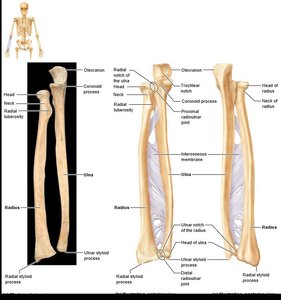
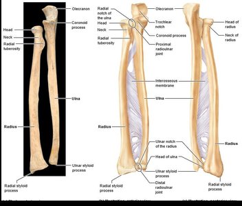
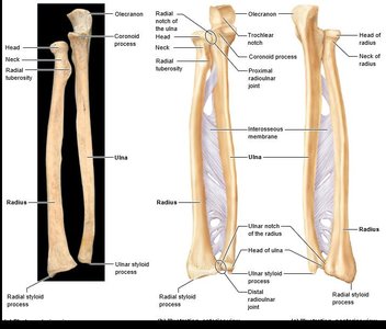
The forearm consists of two parallel bones: the ulna (medial) and radius (lateral).
Proximal ends: Articulate with the humerus and each other.
Distal ends: Articulate with each other at the radioulnar joint.
Interosseous membrane: Connects the radius and ulna along their length.
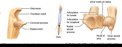
Ulna
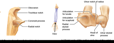
Olecranon and coronoid processes: Grip the trochlea of the humerus, forming a hinge joint.
Radial notch: Articulates with the head of the radius.
Ulnar head: Knoblike distal portion.
Ulnar styloid process: Ligament attachment.
Radius
Head: Articulates with the capitulum of the humerus and radial notch of the ulna.
Radial tuberosity: Anchors the biceps muscle.
Ulnar notch: Articulates with the ulna.
Radial styloid process: Anchors ligaments.
Hand
Bone Structure
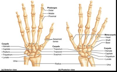
The hand consists of the carpus (wrist), metacarpus (palm), and phalanges (fingers).
Carpus: Eight bones in two rows. Proximal row (lateral to medial): scaphoid, lunate, triquetrum, pisiform. Distal row: trapezium, trapezoid, capitate, hamate. Only scaphoid, lunate, and triquetrum form the wrist joint.
Metacarpus: Five metacarpal bones (I to V from thumb to little finger). Bases articulate with carpals, heads with proximal phalanges.
Phalanges: Fingers numbered I to V starting at thumb (pollex). Digit I has two bones (no middle phalanx); digits II to V have three bones (distal, middle, proximal).
Summary Table: Bones of the Pectoral Girdle and Upper Limb
Body Region | Bones | Location | Markings |
|---|---|---|---|
Pectoral girdle | Clavicle (2), Scapula (2) | Anterior and posterior shoulder | Acromial end, sternal end, spine, acromion, coracoid process |
Upper limb (arm) | Humerus (2) | Arm (shoulder to elbow) | Head, neck, tubercles, epicondyles, trochlea, capitulum |
Forearm | Ulna (2), Radius (2) | Elbow to wrist | Olecranon, coronoid process, radial notch, styloid process |
Hand | Carpals (16), Metacarpals (10), Phalanges (28) | Wrist, palm, fingers | Scaphoid, lunate, triquetrum, pisiform, trapezium, trapezoid, capitate, hamate; base, shaft, head; proximal, middle, distal phalanges |
Pelvic Girdle
Structure and Function
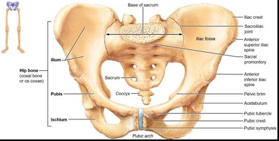
The pelvic girdle, or hip girdle, is formed by two hip bones (coxal bones or os coxae) and the sacrum. It attaches the lower limbs to the axial skeleton with strong ligaments, transmits the weight of the upper body to the lower limbs, and supports pelvic organs.
Mobility: Less than the shoulder joint, but provides more stability.
Coxal Bones
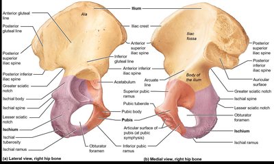
Each coxal bone is formed by the fusion of three bones: ilium, ischium, and pubis.
Ilium: Superior region; auricular surface articulates with sacrum.
Ischium: Posteroinferior part of hip bone.
Pubis: Anterior portion; joins at pubic symphysis.
Acetabulum: Deep socket formed at the point of fusion, receives the head of the femur.
Pelvic Structure and Childbearing
Pelvis Anatomy
The pelvis is formed by the hip bones, sacrum, and coccyx. The female pelvis is wider, shallower, lighter, and rounder than the male’s, adapted for childbearing.
Pelvic brim (pelvic inlet): Continuous oval ridge from pubic crest through arcuate line and sacral promontory.
False pelvis: Superior to pelvic brim.
True pelvis: Inferior to pelvic brim; defines birth canal.
Pelvic outlet: Inferior margin of true pelvis.
The Lower Limb
Bone Structure
The lower limb carries the entire weight of the erect body and is subjected to exceptional forces during jumping or running. It consists of three segments: thigh, leg, and foot.
Femur: Largest and strongest bone in the body; articulates proximally with the acetabulum of the hip and distally with the tibia and patella.
Patella: Sesamoid bone in quadriceps tendon that protects the knee joint.
Tibia: Medial leg bone that receives weight from the femur and transmits it to the foot.
Fibula: Lateral leg bone; not weight bearing, no articulation with femur.
Foot
Tarsus: Seven tarsal bones form the posterior half; body weight carried primarily by talus and calcaneus.
Metatarsals: Five bones (I to V from hallux to little toe); enlarged head of metatarsal I forms the "ball of the foot".
Phalanges: 14 bones of the toes; digit I (hallux) has two bones, digits II to V have three bones.
Arches: Lateral longitudinal, medial longitudinal, and transverse arches allow the foot to bear weight.
Developmental Aspects of the Skeleton
Infant and Adult Skull
Infant skull has more bones than adult skull; bones such as mandible and frontal bones are unfused.
Fontanelles are unossified remnants of fibrous membranes, easing birth and allowing brain growth.
At birth, cranium is large relative to the face; arms and legs grow faster than head and trunk, leading to adult proportions.
Spinal Curvatures and Aging
Primary curvatures of thorax and sacrum are convex at birth, resulting in a C-shaped spine.
Secondary curvatures of cervical and lumbar regions develop as the child grows.
With age, intervertebral discs become thinner, less hydrated, and less elastic, increasing the risk of disc herniation.
Height loss and ossification of costal cartilages are common, leading to a rigid thorax and less efficient gas exchange.
All bones lose mass, increasing fracture risk.
Key Terms and Concepts
Acromioclavicular joint: Joint between the acromion of the scapula and the clavicle.
Glenoid cavity: Shallow socket in the scapula for the head of the humerus.
Trochlea: Hourglass-shaped condyle of the humerus that articulates with the ulna.
Capitulum: Ball-like condyle of the humerus that articulates with the radius.
Epicondyles: Sites of muscle attachment on the humerus.
Interosseous membrane: Fibrous sheet connecting the radius and ulna or tibia and fibula.
Acetabulum: Deep socket in the hip bone for the head of the femur.
Pelvic brim: Edge separating the false and true pelvis.
Sample Exam Questions
The surgical neck is the most frequently fractured portion of the humerus.
The "funny bone" of the elbow is actually the medial epicondyle of the humerus.
The scaphoid and lunate are the two carpals that articulate with the radius to form the wrist joint.
A ring would bear on a phalanx.
Each hip bone is comprised of three separate bones: ilium, ischium, and pubis.
The femur is capable of bearing the greatest physical stress.
The fibula bears no weight in the leg.
Primary curvatures are well developed at birth.
As we age, intervertebral discs become thinner, less elastic, and less hydrated; risk of disc herniation increases.
Additional info:
Tables and images have been recreated and described for clarity and completeness.
Scientific terms have been defined and expanded for academic context.
