 Back
BackThe Appendicular Skeleton: Structure and Function
Study Guide - Smart Notes
 Tailored notes based on your materials, expanded with key definitions, examples, and context.
The Appendicular Skeleton
Overview of the Human Skeleton
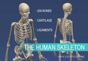
The human skeleton is composed of 206 bones, along with cartilage and ligaments that provide structure, support, and movement. The skeleton is divided into two main parts: the axial skeleton and the appendicular skeleton. The appendicular skeleton includes the limbs and girdles that attach them to the axial skeleton.
Axial Skeleton: Includes the skull, vertebral column, and rib cage.
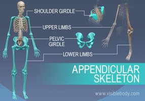
Appendicular Skeleton: Includes the pectoral (shoulder) girdle, pelvic girdle, upper limbs, and lower limbs.
Components of the Appendicular Skeleton
The appendicular skeleton consists of four major regions: the pectoral girdle, upper limbs, pelvic girdle, and lower limbs. These regions are essential for movement and interaction with the environment.
Pectoral Girdle: Composed of the clavicle and scapula, it attaches the upper limbs to the trunk and provides mobility.
Upper Limbs: Includes the humerus, radius, ulna, carpals, metacarpals, and phalanges.
Pelvic Girdle: Formed by the coxal bones (ilium, ischium, pubis), it attaches the lower limbs and supports pelvic organs.
Lower Limbs: Includes the femur, tibia, fibula, tarsals, metatarsals, and phalanges.
Pectoral Girdle
Structure and Function
The pectoral girdle consists of two pairs of bones: the clavicles and scapulae. These bones form a nearly complete circle around the upper trunk and are designed for mobility and flexibility.
Clavicle: The collarbone, mildly S-shaped, articulates with the sternum and scapula. It acts as a brace to push arms laterally and provides muscle attachment points.
Scapula: The shoulder blade, a thin, triangular bone located between ribs 2 and 7. It features the glenoid cavity (shoulder joint), spine, acromion, and coracoid process.
Major Functions:
Attachment points for muscles that move the upper limbs
Allows for a wide range of motion due to shallow and poorly reinforced socket
Upper Limb
Bones and Principal Markings
The upper limb is composed of 30 bones, forming the framework for the arm, forearm, and hand.
Arm: Humerus (longest bone of the upper limb, articulates with scapula, radius, and ulna)
Forearm: Radius and ulna (parallel long bones, articulate with humerus and wrist bones)
Hand: 8 carpal bones (wrist), 5 metacarpal bones (palm), 14 phalanges (fingers)
Key Markings:
Humerus: Head, anatomical neck, surgical neck, tubercles, fossae, condyles, epicondyles
Ulna: Olecranon, coronoid process, styloid process
Radius: Head, ulnar notch, styloid process
Articulation: The elbow joint is formed by the humerus, radius, and ulna, allowing for flexion and extension.
Pelvic Girdle
Structure and Function
The pelvic girdle consists of two hip bones (coxal bones), which unite anteriorly and with the sacrum posteriorly. It forms a complete circle and supports the lower limbs and pelvic organs.
Ilium: Large, flaring bone with iliac crest and spines
Ischium: Postero-inferior part, features ischial tuberosity
Pubis: Anterior part, forms pubic symphysis
Acetabulum: The socket where all three bones join, forming the hip joint.
Pelvic Structure and Childbearing
The female pelvis is wider, shallower, lighter, and rounder than the male pelvis, adapted for childbearing. The pelvic brim separates the false pelvis (abdomen) from the true pelvis (pelvic cavity).
True pelvis: Defines the birth canal
Pelvic outlet: Inferior margin of the true pelvis
Lower Limb
Bones and Principal Markings
The lower limb carries the entire weight of the body and is subjected to exceptional forces. It consists of the thigh, leg, and foot.
Thigh: Femur (largest, strongest bone, articulates with hip and knee)
Leg: Tibia (weight-bearing), fibula (not weight-bearing)
Foot: 26 bones: tarsals (ankle), metatarsals, phalanges
Key Markings:
Femur: Head, neck, trochanters, condyles, epicondyles, patellar surface
Tibia: Condyles, tuberosity, medial malleolus
Fibula: Head, lateral malleolus
Arches of the Foot
The foot has three arches maintained by interlocking bones, ligaments, and tendons, allowing it to bear weight efficiently.
Lateral longitudinal: Low curve elevates lateral part
Medial longitudinal: Arch curves upward
Transverse: Runs obliquely across the foot
Comparative Table: Upper vs Lower Limb Bones
Region | Main Bones | Key Functions |
|---|---|---|
Upper Limb | Humerus, radius, ulna, carpals, metacarpals, phalanges | Manipulation, grasping, movement |
Lower Limb | Femur, tibia, fibula, tarsals, metatarsals, phalanges | Support, locomotion, weight-bearing |
Key Terms and Definitions
Girdle: A set of bones that attach limbs to the axial skeleton
Acetabulum: The socket of the hip joint
Interosseous membrane: A fibrous sheet connecting parallel bones (e.g., radius and ulna; tibia and fibula)
Phalanges: Bones of the fingers and toes
Relevant Equations
While the skeleton itself does not involve equations, understanding bone strength and forces can be related to physics:
Force Transmission: (Force equals mass times acceleration)
Bone Density:
