 Back
BackThe Appendicular Skeleton: Structure, Function, and Variation
Study Guide - Smart Notes
 Tailored notes based on your materials, expanded with key definitions, examples, and context.
The Appendicular Skeleton
Overview of the Appendicular Skeleton
The appendicular skeleton consists of the bones of the limbs and the supporting girdles that connect them to the trunk. It plays a crucial role in movement and locomotion, providing attachment points for muscles and supporting the weight of the body, especially in the lower limbs.
Major Divisions: Pectoral girdles, upper limbs, pelvic girdle, lower limbs
Function: Facilitates movement, supports body weight, and provides leverage for muscles
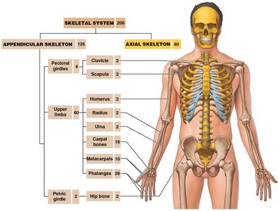
Pectoral Girdles
Structure and Function
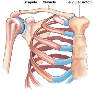
The pectoral (shoulder) girdle connects each arm to the trunk and provides a foundation for arm movement. Each girdle consists of one clavicle and one scapula.
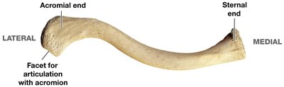
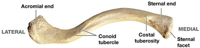
Clavicle: S-shaped bone, connects with the axial skeleton at the manubrium (sternal end) and articulates with the acromion of the scapula (acromial end).
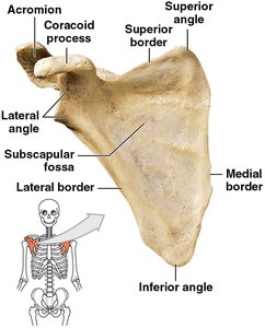
Scapula: Flat, triangular bone located on the posterior side of the rib cage; articulates with the clavicle and humerus.
Mobility: The shoulder is highly mobile but less stable due to the lack of direct bony attachment of the scapula to the axial skeleton.
Clavicle: Surface Features
Conoid tubercle and costal tuberosity: Attachment points for muscles and ligaments.
Sternoclavicular joint: Articulation between clavicle and manubrium of sternum.
Scapula: Surface Features
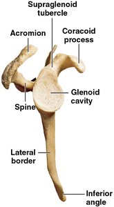
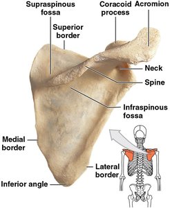
Borders: Superior, medial (vertebral), and lateral (axillary)
Angles: Superior, inferior, lateral
Subscapular fossa: Anterior surface depression
Glenoid cavity: Articulates with the humerus to form the shoulder joint
Processes: Coracoid process (anterior, superior), acromion (posterior, superior), and spine (posterior ridge)
Fossae: Supraspinous (above spine), infraspinous (below spine)
Upper Limbs
Structure and Function
The upper limb consists of the arm (humerus), forearm (ulna and radius), wrist (carpals), and hand (metacarpals and phalanges). These bones facilitate a wide range of movements and dexterity.
Arm (Brachium): Humerus
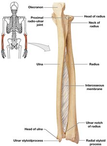
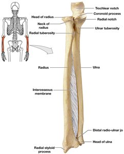
Forearm (Antebrachium): Ulna (medial), Radius (lateral)
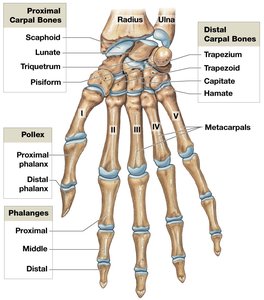
Wrist: Eight carpal bones
Hand: Five metacarpals, 14 phalanges
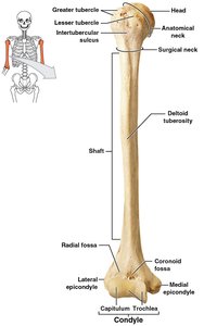
Humerus: Structure and Surface Features
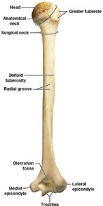
Head: Proximal, articulates with scapula
Greater and lesser tubercles: Muscle attachment sites
Intertubercular sulcus: Groove between tubercles
Anatomical and surgical necks: Anatomical neck is between tubercles and head; surgical neck is a common fracture site
Deltoid tuberosity: Attachment for deltoid muscle
Radial groove: Path for radial nerve
Epicondyles (medial and lateral): Muscle attachment; medial is the "funny bone"
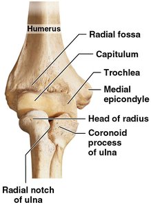
Condyle: Articulates with ulna and radius; includes trochlea (medial) and capitulum (lateral)
Forearm: Ulna and Radius
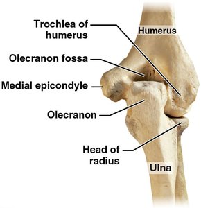
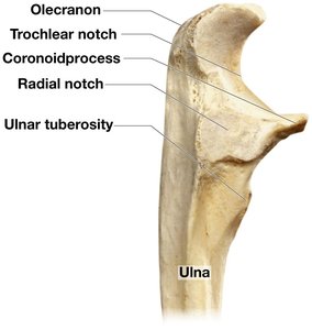
Ulna: Medial bone; olecranon forms elbow point; trochlear notch articulates with humerus; coronoid process fits into humerus during flexion; radial notch for proximal radio-ulnar joint; ulnar styloid process stabilizes wrist.
Radius: Lateral bone; head articulates with capitulum of humerus; radial tuberosity for biceps attachment; ulnar notch for distal radio-ulnar joint; radial styloid process stabilizes wrist.
Interosseous membrane: Connects ulna and radius.
Wrist and Hand
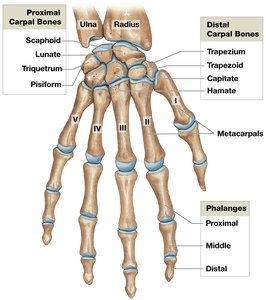
Carpal bones: Eight per wrist (scaphoid, lunate, triquetrum, pisiform, trapezium, trapezoid, capitate, hamate)
Metacarpals: Five per hand, numbered I–V from lateral (thumb) to medial
Phalanges: 14 per hand; thumb (pollex) has two, other fingers have three each
Carpal tunnel syndrome: Compression of the median nerve due to inflammation in the carpal tunnel
Pelvic Girdle
Structure and Function
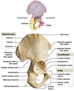
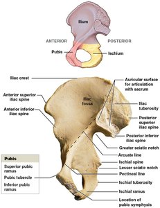
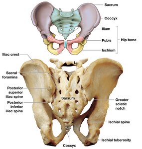
The pelvic girdle attaches the lower limbs to the axial skeleton and supports the weight of the upper body. It consists of two hip bones (coxal bones), each formed by the fusion of the ilium, ischium, and pubis.
Ilium: Superior portion
Ischium: Posterior and inferior portion
Pubis: Anterior and inferior portion
Surface Features of the Hip Bone
Acetabulum: Socket for femur head; formed by all three hip bone components
Obturator foramen: Large opening for nerves and blood vessels
Pubic symphysis: Joint between left and right pubic bones
Iliac crest, iliac fossa, arcuate line: Support and muscle attachment
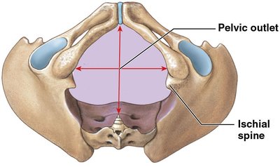
Ischial spine, ischial tuberosity: Muscle and ligament attachment; supports body weight when sitting
The Pelvis
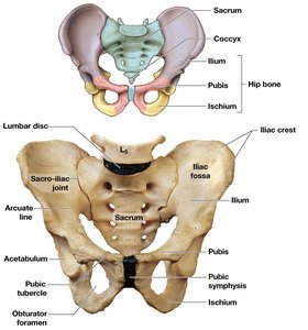
Composed of: Two hip bones, sacrum, coccyx
Sacroiliac joints: Articulation between ilia and sacrum
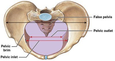
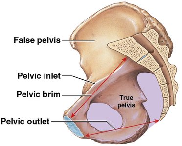
Pelvic cavity: Enclosed by pelvic bones
True (lesser) pelvis: Inferior to pelvic brim
False (greater) pelvis: Superior to pelvic brim
Pelvic inlet and outlet: Boundaries important for childbirth
Perineum: Floor of the pelvic cavity, supports pelvic organs
Sex Differences in the Pelvis
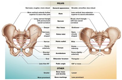
Female pelvis: Broader, smoother, lighter, wider pelvic outlet, triangular obturator foramen, pubic angle ≥ 100°
Male pelvis: Narrower, rougher, more robust, oval obturator foramen, pubic angle ≤ 90°
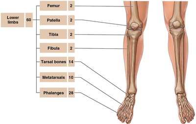
Lower Limbs
Structure and Function
The lower limb bones are specialized for weight bearing and movement. They include the femur, patella, tibia, fibula, tarsals, metatarsals, and phalanges.
Femur: Thigh bone, longest and heaviest in the body
Patella: Kneecap, a sesamoid bone within the quadriceps tendon
Tibia: Medial, weight-bearing bone of the leg
Fibula: Lateral, slender bone of the leg
Tarsals: Seven ankle bones
Metatarsals: Five foot bones
Phalanges: 14 toe bones
Femur: Surface Features
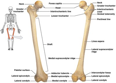
Head: Articulates with acetabulum of hip bone
Fovea capitis: Ligament attachment site
Neck: Connects head to shaft
Greater and lesser trochanters: Muscle attachment
Linea aspera: Posterior ridge for muscle attachment
Epicondyles and condyles: Form part of the knee joint
Patella
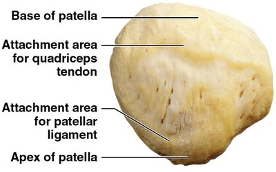
Sesamoid bone: Develops within the quadriceps tendon
Base: Attachment for quadriceps tendon
Apex: Attachment for patellar ligament
Leg: Tibia and Fibula
Tibia: Medial, weight-bearing; medial and lateral condyles articulate with femur; tibial tuberosity for patellar ligament; medial malleolus at ankle
Fibula: Lateral, non-weight-bearing; head articulates with tibia; lateral malleolus at ankle
Interosseous membrane: Connects tibia and fibula
Pott's fracture: Fracture involving both malleoli
Ankle and Foot
Tarsal bones: Talus (articulates with tibia), calcaneus (heel), navicular, cuboid, three cuneiforms
Metatarsals: Five per foot, numbered I–V from medial to lateral
Phalanges: 14 per foot; hallux (great toe) has two, others have three each
Arches: Longitudinal and transverse arches distribute weight and provide support; flatfeet result from loss of these arches
Individual Skeletal Variation
Sex and Age-Related Changes
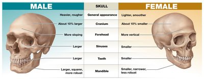
Skeletal features can reveal information about muscle strength, medical history, sex, age, and body size. The female pelvis is modified for childbearing, and age-related changes include fusion of bones and reduction in bone mass.
Relaxin: Hormone that loosens pelvic ligaments during pregnancy
Age-related changes: Fusion of fontanelles, sutures, and epiphyses; reduction in bone mass and density with age
Region/Feature | Event | Age (years) |
|---|---|---|
Bony matrix | Reduction in mineral content; increased osteoporosis risk | Begins 30–45; accelerates after 65 |
Fontanelles (skull) | Closure | By age 2 |
Frontal suture (skull) | Fusion | 2–8 |
Epiphyseal cartilages (long bones) | Fusion | Begins ~3; varies by bone |
Epiphyses (girdles) | Fusion | 14–25 (varies by site) |
Additional info: Bone markings become less prominent with age due to decreased muscle mass and strength. The timing of bone fusion can be used to estimate age in forensic and anthropological contexts.
