 Back
The Appendicular Skeleton: Structure, Function, and Clinical Relevance
Study Guide - Smart Notes
 Tailored notes based on your materials, expanded with key definitions, examples, and context.
The Appendicular Skeleton
Overview of the Appendicular Skeleton
The appendicular skeleton consists of the bones of the limbs and the girdles that attach them to the axial skeleton. It is essential for movement and manipulation of the environment, comprising 126 bones in total.
Pectoral girdles: Attach the upper limbs to the trunk.
Upper extremities: Arms, forearms, wrists, and hands.
Pelvic girdles: Attach the lower limbs to the trunk.
Lower extremities: Thighs, legs, ankles, feet, and toes.
Key Point: The appendicular skeleton is crucial for locomotion and manipulation, providing attachment points for muscles and facilitating a wide range of movements.
The Pectoral Girdle
Structure and Articulation
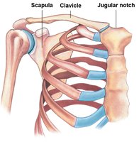
The pectoral (shoulder) girdle connects the upper limbs to the axial skeleton. Each arm articulates with the trunk at this girdle, which consists of the clavicles and scapulae.
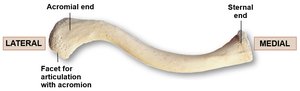
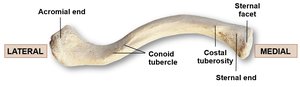
Clavicles: S-shaped bones that connect the sternum to the scapulae.
Scapulae: Flat, triangular bones that provide attachment for muscles and articulate with the humerus.
Clavicle
The clavicle acts as a strut to keep the upper limb away from the thorax, allowing for maximum range of motion.
Acromial end: Articulates with the acromion of the scapula.
Sternal end: Articulates with the manubrium of the sternum.
Scapula
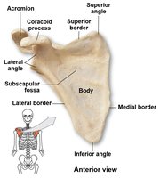
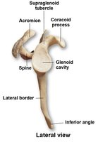
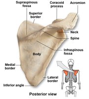
The scapula is a broad, flat bone with several important anatomical features for muscle attachment and articulation with the humerus and clavicle.
Glenoid cavity: Articulates with the head of the humerus.
Coracoid process: Attachment for ligaments and tendons.
Acromion: Articulates with the clavicle.
Scapular spine: Prominent ridge on the posterior surface.
Fossae: Subscapular (anterior), supraspinous (above spine), and infraspinous (below spine).
Upper Extremities
Regions and Bones
The upper extremity is divided into several regions, each with specialized bones for movement and manipulation.
Brachium: Humerus
Antebrachium: Radius and ulna
Wrist: Carpals
Hand: Metacarpals and phalanges
Humerus
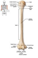
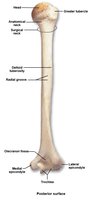
The humerus is the only bone of the arm, articulating proximally with the scapula and distally with the radius and ulna.
Greater and lesser tubercles: Muscle attachment sites.
Intertubercular groove: Passage for the tendon of the biceps brachii.
Deltoid tuberosity: Attachment for the deltoid muscle.
Epicondyles: Medial and lateral, for muscle attachment.
Condyle: Articulates with forearm bones.
Fossae: Coronoid, olecranon, and radial for accommodating processes of the ulna and radius during movement.
Elbow Joint
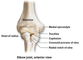
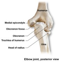
The elbow is a hinge joint formed by the articulation of the humerus with the radius and ulna.
Trochlea: Articulates with the ulna.
Capitulum: Articulates with the radius.
Olecranon: Point of the elbow, part of the ulna.
Ulna and Radius
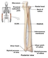
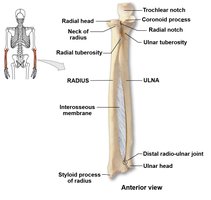
The ulna is the medial bone of the forearm, while the radius is lateral. They articulate with each other at the proximal and distal radioulnar joints and are connected by an interosseous membrane.
Ulna: Olecranon, trochlear notch, coronoid process, ulnar head, styloid process.
Radius: Radial head, radial tuberosity, ulnar notch, styloid process.
Carpal Bones, Metacarpals, and Phalanges
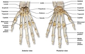
The wrist contains eight carpal bones, while the hand consists of five metacarpals and fourteen phalanges. The thumb (pollex) has only two phalanges, while the other digits have three each.
Carpals: Scaphoid, lunate, triquetrum, pisiform (proximal); trapezium, trapezoid, capitate, hamate (distal).
Metacarpals: Numbered I-V from lateral (thumb) to medial.
Phalanges: Proximal, middle, and distal (except thumb).
The Pelvic Girdle
Structure and Features
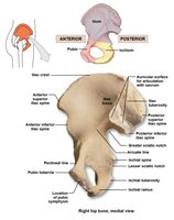
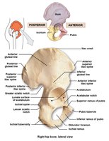
The pelvic girdle consists of two hip bones (coxal bones), each formed by the fusion of the ilium, ischium, and pubis. The acetabulum is the socket for the femoral head.
Ilium: Superior portion, includes iliac crest and fossa.
Ischium: Posterior-inferior portion, includes ischial spine and tuberosity.
Pubis: Anterior portion, includes pubic symphysis and superior ramus.
Greater sciatic notch: Passage for the sciatic nerve.
Auricular surface: Articulates with the sacrum.
Pelvis
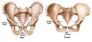
The pelvis is formed by the two hip bones, sacrum, and coccyx. It is divided into the true (lesser) and false (greater) pelvis. The female pelvis is generally broader and lighter than the male pelvis, with a wider pelvic inlet and outlet.
Lower Extremities
Regions and Bones
The lower extremity is specialized for weight-bearing and locomotion, consisting of the femur, patella, tibia, fibula, tarsals, metatarsals, and phalanges.
Thigh: Femur
Kneecap: Patella
Leg: Tibia (medial) and fibula (lateral)
Ankle: Tarsals
Foot: Metatarsals and phalanges
Femur
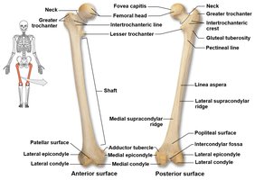
The femur is the longest and strongest bone in the body, articulating with the hip and tibia.
Femoral head: Articulates with the acetabulum.
Neck: Connects head to shaft.
Greater and lesser trochanters: Muscle attachment sites.
Linea aspera: Posterior ridge for muscle attachment.
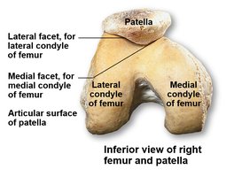
Condyles and epicondyles: Form part of the knee joint.
Patella
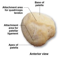
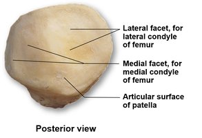
The patella is a sesamoid bone embedded in the quadriceps tendon, protecting the knee joint and improving leverage for the quadriceps muscle.
Base: Attachment for quadriceps tendon.
Apex: Attachment for patellar ligament.
Tibia and Fibula
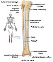
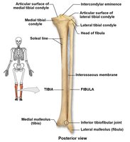
The tibia is the main weight-bearing bone of the leg, while the fibula provides lateral stability and muscle attachment.
Tibia: Medial and lateral condyles, tibial tuberosity, anterior margin, medial malleolus.
Fibula: Head, lateral malleolus.
Interosseous membrane: Connects tibia and fibula.
Tarsal Bones
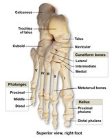
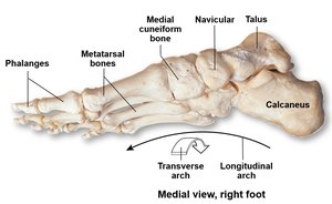
The tarsus consists of seven bones forming the ankle and proximal foot. The talus transmits weight from the tibia, and the calcaneus forms the heel.
Talus: Superior tarsal bone, articulates with tibia and fibula.
Calcaneus: Largest tarsal bone, forms the heel.
Navicular, cuboid, cuneiforms (medial, intermediate, lateral): Form the arch and structure of the foot.
Metatarsals and Phalanges
The foot contains five metatarsals and fourteen phalanges. The great toe (hallux) has only two phalanges, while the other toes have three each.
Metatarsals: Numbered I-V from medial (hallux) to lateral.
Phalanges: Proximal, middle, and distal (except hallux).
Male and Female Bone Comparison
Pelvis, Skull, and Other Skeletal Elements
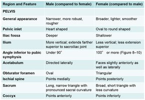
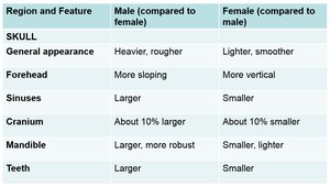
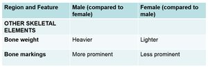
There are notable differences between male and female skeletons, especially in the pelvis and skull, which are important for forensic and clinical identification.
Region and Feature | Male (compared to female) | Female (compared to male) |
|---|---|---|
Pelvis: General appearance | Narrower, more robust, rougher | Broader, lighter, smoother |
Pelvic inlet | Heart shaped | Oval to round shaped |
Iliac fossa | Deeper | Shallower |
Angle inferior to pubic symphysis | Under 90° | 100° or more |
Acetabulum | Directed laterally | Faces slightly anteriorly as well as laterally |
Obturator foramen | Oval | Triangular |
Ischial spine | Points medially | Points posteriorly |
Sacrum | Long, narrow triangle with pronounced sacral curvature | Broad, short triangle with less curvature |
Coccyx | Points anteriorly | Points inferiorly |
Age-Related Changes in the Skeleton
General Skeletal Changes
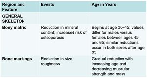
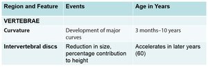
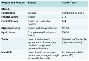
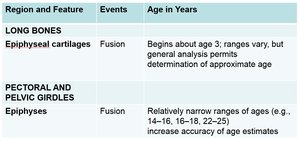
With age, the skeleton undergoes several changes, including reduction in bone mass, changes in bone markings, and fusion of growth plates. These changes can be used to estimate age in forensic and clinical settings.
Region and Feature | Events | Age in Years |
|---|---|---|
General Skeleton: Bony matrix | Reduction in mineral content; increased risk of osteoporosis | Begins at age 30–45 |
Bone markings | Reduction in size, roughness | Gradual with age |
Vertebrae: Curvature | Development of major curves | 3 months–10 years |
Intervertebral discs | Reduction in size, height | Accelerates after age 60 |
Skull: Fontanelles | Closure | By age 2 |
Long bones: Epiphyseal cartilages | Fusion | Begins about age 3 |
Pectoral and pelvic girdles: Epiphyses | Fusion | Ranges 14–25 years |
Additional info: Age-related changes are important for understanding osteoporosis, growth, and forensic identification. Differences in male and female skeletons are especially relevant in anthropology and medicine.
