 Back
The Appendicular Skeleton: Structure, Function, and Clinical Correlates
Study Guide - Smart Notes
 Tailored notes based on your materials, expanded with key definitions, examples, and context.
The Appendicular Skeleton
Overview of the Appendicular Skeleton
The appendicular skeleton consists of the bones of the limbs and the girdles that attach them to the axial skeleton. It is essential for movement and manipulation of the environment. The appendicular skeleton is divided into the pectoral (shoulder) girdle, upper limbs, pelvic (hip) girdle, and lower limbs.
Pectoral girdle: Attaches the upper limbs to the trunk.
Pelvic girdle: Attaches the lower limbs to the trunk.
Upper and lower limbs differ in function but share a similar structural plan.
The Pectoral Girdle
Structure and Function
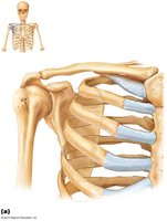
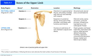
The pectoral girdle consists of the clavicle and scapula, providing attachment points for muscles and allowing a wide range of motion for the upper limb. Only the clavicle articulates directly with the axial skeleton, while the scapula is highly mobile.
Clavicle: Extends horizontally across the superior thorax; sternal end articulates with the manubrium, acromial end with the scapula.
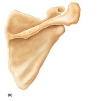
Scapula: Lies on the dorsal surface of the rib cage, between ribs 2–7; has three borders (superior, medial, lateral) and three angles (lateral, superior, inferior).
Glenoid cavity: Shallow socket for the humerus, allowing flexibility but less stability.
The Upper Limb
Bone Groups and Key Features
Each upper limb contains 30 bones, grouped into the arm, forearm, and hand. These bones provide structure, leverage, and articulation for complex movements.
Arm: Humerus is the sole bone, articulating with the scapula and forearm bones.
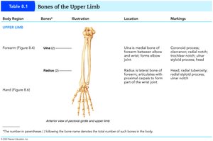
Forearm: Consists of the radius and ulna, which articulate with each other at proximal and distal radioulnar joints.
Hand: Includes the carpal bones (wrist), metacarpals (palm), and phalanges (fingers).
Articulations of the Upper Limb
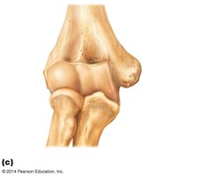
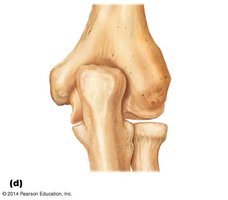
The elbow joint is a complex hinge joint involving the humerus, radius, and ulna. The radius and ulna are connected by an interosseous membrane, allowing pronation and supination of the forearm.
Humerus: Features include the head, anatomical and surgical necks, greater and lesser tubercles, and condyles for articulation with the forearm.
Radius and Ulna: The ulna forms the elbow's hinge with the humerus; the radius allows rotation of the hand.
The Pelvic Girdle
Structure and Function
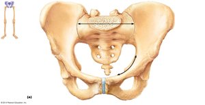
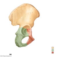
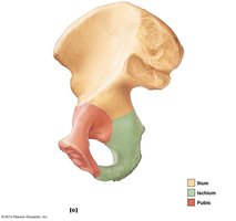
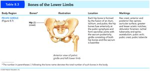
The pelvic girdle consists of two hip bones (os coxae), each formed by the fusion of the ilium, ischium, and pubis. It attaches the lower limbs to the axial skeleton and supports the weight of the upper body.
Ilium: Superior, flaring portion of the hip bone.
Ischium: Posteroinferior part of the hip bone.
Pubis: Anterior portion of the hip bone.
Acetabulum: Deep socket for the femoral head, providing stability for the hip joint.
Comparison of Male and Female Pelves
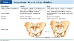
The male and female pelves differ in structure due to functional requirements, such as childbirth in females. These differences are evident in the shape, size, and angle of the pelvic bones.
Characteristic | Female | Male |
|---|---|---|
General structure | Adapted for childbirth; lighter, thinner, broader | Heavier, thicker, more prominent markings |
Pelvic inlet | Broader, more oval | Narrower, more heart-shaped |
Pubic arch angle | Broader (80–90°) | More acute (50–60°) |
The Lower Limb
Bone Groups and Key Features
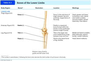
The lower limb is specialized for weight-bearing and locomotion. It consists of the thigh, leg, and foot, each with distinct bones and articulations.
Thigh: Femur is the largest and strongest bone in the body.
Leg: Tibia (medial, weight-bearing) and fibula (lateral, stabilizing).
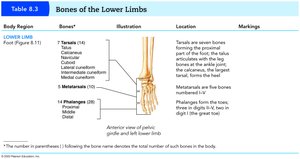
Foot: Tarsals (ankle), metatarsals (sole), and phalanges (toes).
Disorders of the Appendicular Skeleton
Common Disorders
Several congenital and acquired disorders can affect the appendicular skeleton, impacting mobility and quality of life.
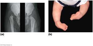
Hip dysplasia: The head of the femur slips out of the acetabulum, often present at birth.
Clubfoot: Soles of the feet turn medially, a congenital condition.
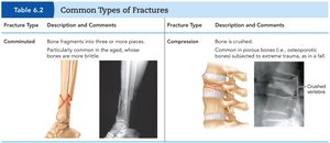
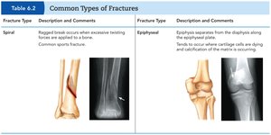
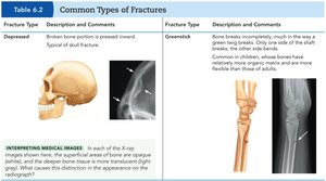
Bone fractures: Various types, including comminuted, compression, spiral, epiphyseal, depressed, and greenstick fractures.
Common Types of Fractures
Fracture Type | Description and Comments |
|---|---|
Comminuted | Bone fragments into three or more pieces; common in aged, brittle bones. |
Compression | Bone is crushed; common in porous bones subjected to trauma. |
Spiral | Ragged break from excessive twisting; common in sports injuries. |
Epiphyseal | Epiphysis separates from diaphysis along growth plate; occurs where cartilage cells are dying. |
Depressed | Broken bone portion pressed inward; typical of skull fractures. |
Greenstick | Bone breaks incompletely, like a green twig; more common in children. |
Additional info: The appendicular skeleton is crucial for movement, support, and interaction with the environment. Disorders and injuries can significantly impact function, highlighting the importance of understanding its anatomy and clinical relevance.
