Back
BackThe Appendicular Skeleton: Structure, Function, and Development
Study Guide - Smart Notes
 Tailored notes based on your materials, expanded with key definitions, examples, and context.
The Appendicular Skeleton
Overview of the Appendicular Skeleton
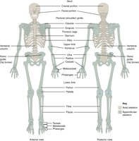
The appendicular skeleton includes the bones of the upper and lower limbs, as well as the girdles that attach them to the axial skeleton. It is essential for movement and manipulation of the environment.
Main components: Pectoral girdle, upper limbs, pelvic girdle, lower limbs
Function: Facilitates locomotion and interaction with surroundings
Attachment: Girdles anchor limbs to the axial skeleton
The Pectoral Girdle
Structure and Function
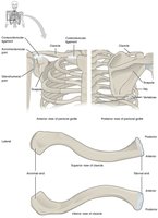
The pectoral girdle consists of the clavicle and scapula, which attach the upper limb to the trunk. This structure allows for a wide range of motion at the shoulder.
Clavicle: S-shaped bone that connects the arm to the trunk at the sternum
Scapula: Flat, triangular bone providing attachment for muscles and articulation with the humerus
Function: Supports arm movement and stabilizes the shoulder
Anatomy of the Scapula
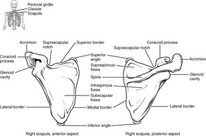
The scapula has several important landmarks for muscle attachment and articulation.
Acromion: Articulates with the clavicle
Glenoid cavity: Articulates with the head of the humerus
Spine, coracoid process, borders, and angles: Serve as muscle attachment sites
Bones of the Upper Limb
Divisions and Major Bones
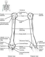
The upper limb is divided into the arm, forearm, and hand, each with distinct bones and joints.
Arm: Humerus (single bone)
Forearm: Radius (lateral), Ulna (medial)
Hand: Carpals (wrist), Metacarpals (palm), Phalanges (fingers)
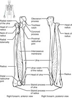
Forearm Anatomy
The radius and ulna are connected by an interosseous membrane and articulate with each other at proximal and distal radioulnar joints.
Radius: Lateral bone, important for wrist movement
Ulna: Medial bone, forms the elbow joint with the humerus
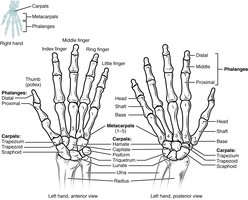
Bones of the Hand
The hand consists of the carpal bones (wrist), metacarpals (palm), and phalanges (fingers).
Carpals: Eight bones arranged in two rows
Metacarpals: Five bones forming the palm
Phalanges: Fourteen bones in the fingers (three per finger, two in the thumb)
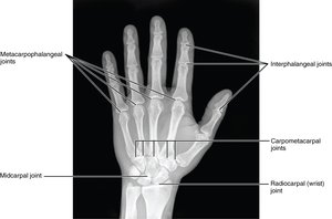
Radiographic Anatomy of the Hand
X-rays reveal the arrangement of the hand's bones and joints, which are crucial for diagnosing fractures and other conditions.
The Carpal Tunnel
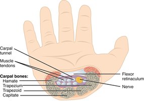
The carpal tunnel is a passageway on the anterior side of the wrist, formed by carpal bones and the flexor retinaculum. It allows passage of tendons and the median nerve into the hand.
Clinical relevance: Compression of the median nerve leads to carpal tunnel syndrome
Hand Grip Mechanics
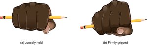
During a firm grip, the fourth and fifth metacarpals move anteriorly, increasing contact and grip strength.
Common Upper Limb Fractures
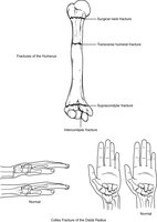
Fractures can occur at various sites in the upper limb, with the distal radius (Colles fracture) being the most common in the forearm.
Surgical neck of humerus: Common fracture site in elderly
Colles fracture: Distal radius fracture, often from falls
The Pelvic Girdle and Pelvis
Structure and Function
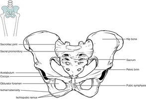
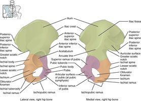
The pelvic girdle is formed by the hip bones, which attach the lower limbs to the axial skeleton. The pelvis includes the hip bones, sacrum, and coccyx.
Function: Supports body weight, protects pelvic organs, and provides attachment for lower limbs
Regions of the Hip Bone
The adult hip bone is composed of three fused regions: ilium, ischium, and pubis.
Ilium: Superior, fan-shaped portion
Ischium: Posteroinferior portion
Pubis: Anteromedial portion
Ligaments of the Pelvis
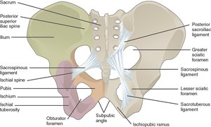
Strong ligaments stabilize the pelvis and form important foramina for neurovascular structures.
Posterior sacroiliac ligament: Supports sacroiliac joint
Sacrospinous and sacrotuberous ligaments: Form greater and lesser sciatic foramina
Sex Differences in the Pelvis
The female pelvis is adapted for childbirth, being broader and more spacious than the male pelvis.
Female pelvis: Broader, larger subpubic angle, rounder pelvic brim, wider lesser pelvic cavity
Male pelvis: Narrower, heavier, smaller subpubic angle
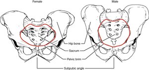
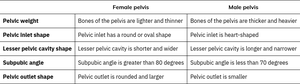
Table: Comparison of Female and Male Pelvis
Female pelvis | Male pelvis | |
|---|---|---|
Pelvic weight | Bones of the pelvis are lighter and thinner | Bones of the pelvis are thicker and heavier |
Pelvic inlet shape | Pelvic inlet has a round or oval shape | Pelvic inlet is heart-shaped |
Lesser pelvic cavity shape | Lesser pelvic cavity is shorter and wider | Lesser pelvic cavity is longer and narrower |
Subpubic angle | Subpubic angle is greater than 80 degrees | Subpubic angle is less than 70 degrees |
Pelvic outlet shape | Pelvic outlet is rounded and larger | Pelvic outlet is smaller |
Bones of the Lower Limb
Divisions and Major Bones
The lower limb is divided into the thigh, leg, and foot, each with specialized bones for weight-bearing and movement.
Thigh: Femur (single bone)
Leg: Tibia (medial, weight-bearing), Fibula (lateral, slender)
Foot: Tarsals (ankle), Metatarsals (mid-foot), Phalanges (toes)
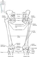
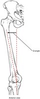
The Q-Angle
The Q-angle measures the lateral deviation of the femur from the vertical axis of the tibia. It is typically larger in females due to a wider pelvis.
Clinical relevance: Increased Q-angle can predispose to knee injuries
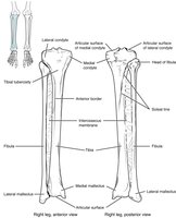
Leg Bones: Tibia and Fibula
The tibia is the main weight-bearing bone of the leg, while the fibula provides lateral stability and muscle attachment.
Tibia: Medial, larger, supports body weight
Fibula: Lateral, slender, not weight-bearing
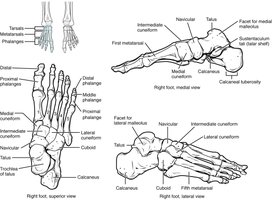
Bones of the Foot
The foot is structured for support and movement, with tarsal bones forming the ankle, metatarsals the mid-foot, and phalanges the toes.
Tarsals: Seven bones, including the calcaneus (heel) and talus (ankle joint)
Metatarsals: Five bones
Phalanges: Fourteen bones (three per toe, two in the big toe)
Development of the Appendicular Skeleton
Embryonic Limb Buds
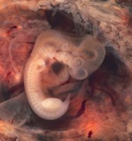
Limb buds appear in the embryo by the end of the seventh week, marking the beginning of appendicular skeleton development.
Ossification Centers and Congenital Deformities
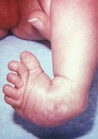
Primary and secondary ossification centers form in the developing bones. Congenital deformities, such as clubfoot, can occur but are often treatable.
Primary ossification: Begins in the diaphysis (shaft) of long bones
Secondary ossification: Occurs in the epiphyses (ends) after birth
Clubfoot: Common congenital deformity, usually correctable