 Back
BackThe Appendicular Skeleton: Structure, Markings, and Identification
Study Guide - Smart Notes
 Tailored notes based on your materials, expanded with key definitions, examples, and context.
The Appendicular Skeleton
Overview of the Appendicular Skeleton
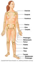
The appendicular skeleton consists of the bones of the limbs and the girdles that attach them to the axial skeleton. It is essential for movement and manipulation of the environment.
Pectoral girdle: Attaches the upper limbs to the trunk.
Pelvic girdle: Attaches the lower limbs to the trunk.
Upper limbs: Includes the arm, forearm, and hand.
Lower limbs: Includes the thigh, leg, and foot.
Pectoral Girdle
Clavicle and Scapula
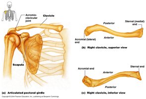
The pectoral girdle is composed of the clavicle and scapula, which provide attachment points for muscles and facilitate arm movement.
Clavicle: S-shaped bone connecting the sternum to the scapula.
Scapula: Flat, triangular bone located on the posterior thorax.
Acromioclavicular joint: The joint between the acromion of the scapula and the clavicle.
Bone markings: Sternal end, acromial end, and various ridges and fossae.
Upper Limb
Humerus
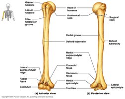
The humerus is the bone of the upper arm, articulating with the scapula at the shoulder and with the radius and ulna at the elbow.
Head: Articulates with the glenoid cavity of the scapula.
Greater and lesser tubercles: Sites for muscle attachment.
Deltoid tuberosity: Attachment for the deltoid muscle.
Epicondyles: Medial and lateral projections for muscle and ligament attachment.
Radius and Ulna
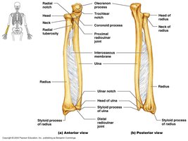
The radius and ulna are the bones of the forearm. The radius is lateral (thumb side), and the ulna is medial (little finger side).
Olecranon process: Prominent projection of the ulna forming the elbow.
Radial tuberosity: Attachment for the biceps brachii muscle.
Styloid processes: Distal projections for ligament attachment.
Interosseous membrane: Connects the radius and ulna along their length.
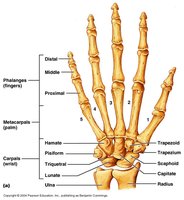
Hand: Carpals, Metacarpals, and Phalanges
The hand consists of the wrist (carpals), palm (metacarpals), and fingers (phalanges).
Carpals: Eight small bones forming the wrist.
Metacarpals: Five bones forming the palm.
Phalanges: Fourteen bones forming the fingers (proximal, middle, distal).
Mnemonic: "Sally Left The Party To Take Cathy Home" helps remember the carpal bones: Scaphoid, Lunate, Triquetrum, Pisiform, Trapezium, Trapezoid, Capitate, Hamate.
Pelvic Girdle
Ilium, Ischium, and Pubis
The pelvic girdle is formed by the fusion of three bones: ilium, ischium, and pubis. It supports the weight of the upper body and protects pelvic organs.
Ilium: Largest, superior bone of the pelvis.
Ischium: Inferior, posterior bone.
Pubis: Anterior bone forming the pubic symphysis.
Lower Limb
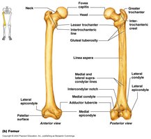
Femur
The femur is the longest and strongest bone in the body, forming the thigh.
Head: Articulates with the acetabulum of the pelvis.
Neck: Connects the head to the shaft.
Greater and lesser trochanters: Muscle attachment sites.
Condyles and epicondyles: Articulate with the tibia and patella.
Tibia and Fibula
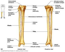
The tibia and fibula are the bones of the lower leg. The tibia is medial and weight-bearing, while the fibula is lateral and slender.
Tibial tuberosity: Attachment for the patellar ligament.
Medial and lateral malleoli: Distal projections forming the ankle.
Interosseous membrane: Connects the tibia and fibula.
Patella
The patella, or kneecap, is a sesamoid bone embedded in the tendon of the quadriceps femoris muscle. It protects the knee joint and improves leverage of the thigh muscles.
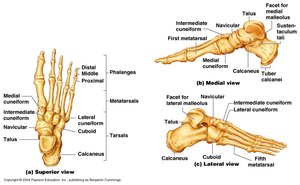
Foot: Tarsals, Metatarsals, and Phalanges
The foot consists of the ankle (tarsals), instep (metatarsals), and toes (phalanges).
Tarsals: Seven bones forming the ankle, including the talus and calcaneus.
Metatarsals: Five bones forming the instep.
Phalanges: Fourteen bones forming the toes (proximal, middle, distal).
Mnemonic: "Today’s Lab Exercises" may refer to learning the bones and their markings in both articulated and disarticulated forms.
Bone Markings and Identification
Bone Markings
Bone markings are anatomical features on bones that serve as sites for muscle attachment, passage of nerves and blood vessels, and articulation with other bones.
Examples: Tuberosity, crest, trochanter, condyle, epicondyle, fossa, foramen.
Application: Identifying bone markings is essential for understanding muscle and ligament attachments and joint function.
Right vs. Left Bones
Determining whether a bone is from the right or left side of the body involves examining anatomical landmarks and orientation.
Example: The head of the humerus faces medially, while the deltoid tuberosity is lateral.
Application: Correct identification is crucial for anatomical study and clinical practice.
Summary Table: Major Bones of the Appendicular Skeleton
Region | Main Bones | Key Markings |
|---|---|---|
Pectoral Girdle | Clavicle, Scapula | Acromion, Sternal end, Spine |
Upper Limb | Humerus, Radius, Ulna | Head, Tuberosity, Epicondyle, Olecranon |
Hand | Carpals, Metacarpals, Phalanges | Proximal, Middle, Distal |
Pelvic Girdle | Ilium, Ischium, Pubis | Crest, Tuberosity, Symphysis |
Lower Limb | Femur, Tibia, Fibula, Patella | Head, Neck, Condyle, Tuberosity |
Foot | Tarsals, Metatarsals, Phalanges | Talus, Calcaneus, Cuneiforms |
Lab Exercise Guidance
Studying Articulated and Disarticulated Bones
In laboratory exercises, students should learn to identify bones and their markings in both articulated (connected) and disarticulated (separated) forms.
Articulated skeleton: Bones joined together as in the living body.
Disarticulated skeleton: Bones separated for individual study.
Learning Bone Markings
Focus on recognizing and naming bone markings, as these are critical for understanding anatomical relationships and functions.
Practice: Use models, diagrams, and hands-on examination to reinforce learning.
----------------------------------------
