 Back
BackThe Auditory System and Balance: Structure, Function, and Pathways
Study Guide - Smart Notes
 Tailored notes based on your materials, expanded with key definitions, examples, and context.
The Ear: Hearing and Balance
Overview of the Auditory and Equilibrium Systems
The ear is responsible for both hearing (audition) and balance (equilibrium). These functions are mediated by specialized mechanoreceptors located in the internal ear. Although the organs for hearing and equilibrium are structurally connected, their receptors respond to different stimuli and are activated independently.
Hearing: Detection of sound waves and their conversion into neural signals.
Equilibrium: Detection of head position and movement to maintain balance.
Sound: Physical Properties and Perception
Nature of Sound Waves
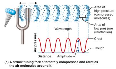
Sound is produced by vibrating objects, which create alternating areas of high and low pressure in the surrounding air. These pressure waves travel through the air and are perceived as sound when they reach the ear.
Wavelength: The distance between two consecutive crests or troughs of a sound wave.
Amplitude: The height of the wave, which determines the loudness of the sound.
Frequency: The number of waves that pass a given point per second (measured in Hertz, Hz); determines the pitch of the sound.
Pitch and Loudness
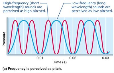
Pitch: Determined by the frequency of the sound wave. High-frequency waves are perceived as high-pitched sounds, while low-frequency waves are perceived as low-pitched sounds.
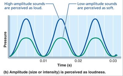
Loudness: Determined by the amplitude of the sound wave. Larger amplitudes are perceived as louder sounds.
Anatomy of the Ear
Three Major Regions of the Ear
The ear is divided into three main regions, each with distinct structures and functions:
External (Outer) Ear: Involved in hearing only.
Middle Ear (Tympanic Cavity): Involved in hearing only.
Internal (Inner) Ear: Involved in both hearing and equilibrium.
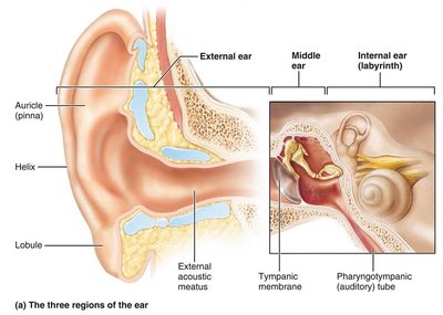
External Ear
Auricle (Pinna): Shell-shaped structure that funnels sound waves into the auditory canal.
External Acoustic Meatus (Auditory Canal): Short, curved tube lined with skin, hairs, sebaceous glands, and ceruminous (earwax) glands; transmits sound waves to the tympanic membrane.
Tympanic Membrane (Eardrum): Thin, translucent membrane that vibrates in response to sound and transfers sound energy to the middle ear bones.
Middle Ear
The middle ear is an air-filled cavity within the temporal bone, containing the auditory ossicles and the pharyngotympanic (auditory) tube.
Auditory Ossicles: Three small bones (malleus, incus, stapes) that transmit and amplify vibrations from the tympanic membrane to the oval window of the inner ear.
Pharyngotympanic (Auditory) Tube: Connects the middle ear to the nasopharynx, equalizing pressure across the tympanic membrane.
Muscles: Tensor tympani and stapedius muscles contract reflexively to protect the inner ear from loud sounds.
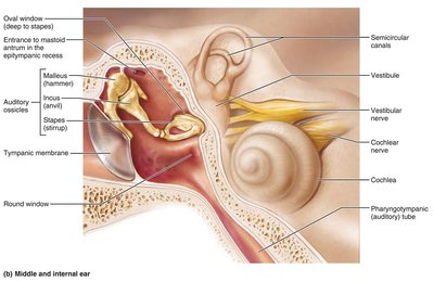
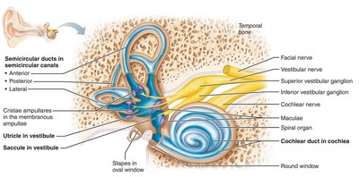
Internal Ear (Labyrinth)
The internal ear consists of a bony labyrinth filled with perilymph and a membranous labyrinth filled with endolymph. It is divided into three main regions:
Vestibule: Central cavity containing the saccule and utricle, which house equilibrium receptors (maculae).
Semicircular Canals: Three canals oriented in different planes, each containing a semicircular duct and an ampulla with the crista ampullaris (receptor for rotational acceleration).
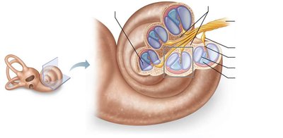
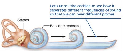
Cochlea: Spiral-shaped organ responsible for hearing, containing the cochlear duct (scala media) and the spiral organ (organ of Corti).
Cochlea: Structure and Function
Anatomy of the Cochlea
The cochlea is a spiral, conical, bony chamber that contains the cochlear duct, which houses the spiral organ (organ of Corti). The cochlear cavity is divided into three chambers:
Scala Vestibuli: Contains perilymph; abuts the oval window.
Scala Media (Cochlear Duct): Contains endolymph; houses the spiral organ.
Scala Tympani: Contains perilymph; terminates at the round window.
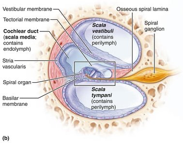
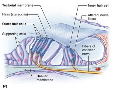
Spiral Organ (Organ of Corti)
The spiral organ is the receptor organ for hearing, containing inner and outer hair cells sandwiched between the tectorial and basilar membranes. The cochlear branch of the vestibulocochlear nerve (VIII) transmits auditory information to the brain.
Summary Table: Internal Ear Structures and Functions
Bony Labyrinth | Membranous Labyrinth | Function | Receptor Region |
|---|---|---|---|
Semicircular canals | Semicircular ducts | Equilibrium: rotational (angular) acceleration | Crista ampullaris |
Vestibule | Utricle and saccule | Equilibrium: head position relative to gravity, linear acceleration | Macula |
Cochlea | Cochlear duct (scala media) | Hearing | Spiral organ |
Pathway of Sound Waves Through the Ear
Transmission of Sound
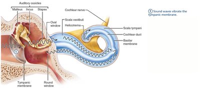
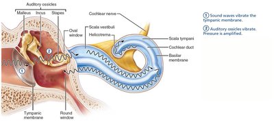
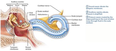
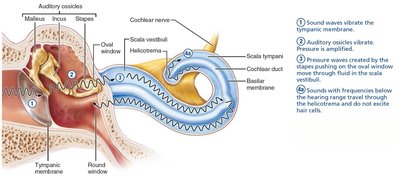
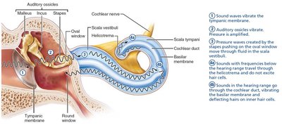
Sound waves enter the external acoustic meatus and strike the tympanic membrane, causing it to vibrate.
Vibrations are transferred to the auditory ossicles, which amplify the pressure and transmit it to the oval window.
The stapes at the oval window creates pressure waves in the perilymph of the scala vestibuli.
Waves with frequencies below the threshold of hearing travel through the helicotrema and do not excite hair cells.
Sounds in the hearing range travel through the cochlear duct, vibrating the basilar membrane and deflecting hairs on inner hair cells.
Resonance of the Basilar Membrane
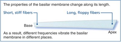
The basilar membrane varies in width and stiffness along its length, allowing it to resonate at different frequencies at different locations:
Base (near oval window): Short, stiff fibers resonate with high-frequency waves.
Apex (far from oval window): Long, floppy fibers resonate with low-frequency waves.

Sound Transduction and Hair Cell Function
Mechanism of Sound Transduction
Movement of the basilar membrane causes deflection of the stereocilia on inner hair cells, which are embedded in the tectorial membrane. This mechanical deflection opens or closes mechanically gated ion channels, leading to changes in the hair cell's membrane potential and neurotransmitter release.
Bending toward the tallest stereocilia opens K+ and Ca2+ channels, causing depolarization.
Bending toward the shortest stereocilia closes channels, causing hyperpolarization.
Role of Inner and Outer Hair Cells
Inner Hair Cells: Primary sensory receptors for hearing; send auditory information to the brain.
Outer Hair Cells: Modulate the responsiveness of the basilar membrane, amplifying or protecting inner hair cells from damage.
Auditory Pathway to the Brain
Neural Pathway
Auditory information is transmitted from the cochlear receptors (inner hair cells) to the cerebral cortex via the cochlear branch of the vestibulocochlear nerve (VIII). The pathway includes several relay stations:
Spiral ganglion
Cochlear nuclei (medulla)
Superior olivary nucleus (pons-medulla)
Lateral lemniscus (tract)
Inferior colliculus (midbrain)
Medial geniculate nucleus (thalamus)
Primary auditory cortex
Auditory Processing
Pitch Perception: Determined by the position of hair cells stimulated along the basilar membrane.
Loudness Detection: Determined by the frequency of action potentials generated by hair cells.
Sound Localization: Depends on the relative intensity and timing of sound waves reaching both ears.
Equilibrium: Static and Dynamic Balance
Vestibular Apparatus
The vestibular apparatus includes the equilibrium receptors in the semicircular canals and vestibule. It monitors both static and dynamic equilibrium:
Vestibular Receptors (Maculae): Monitor static equilibrium (head position relative to gravity and linear acceleration).
Semicircular Canal Receptors (Cristae Ampullares): Monitor dynamic equilibrium (rotational movements).
Maculae
Maculae are sensory receptor organs located in the saccule and utricle. They contain hair cells with stereocilia and a kinocilium, embedded in an otolith membrane studded with calcium carbonate crystals (otoliths). Maculae respond to linear acceleration and head position changes.
Cristae Ampullares
The crista ampullaris is the receptor for rotational acceleration, located in the ampulla of each semicircular canal. Hair cells extend into a gel-like mass called the ampullary cupula. Rotational movements cause endolymph to move, bending the hair cells and altering the rate of nerve impulses sent to the brain.
Equilibrium Pathway to the Brain
Equilibrium information is sent to reflex centers in the brainstem and cerebellum, allowing for rapid adjustments to maintain balance. Input is integrated from vestibular, visual, and somatic receptors.
Clinical Application: Deafness and Cochlear Implants
Sensorineural Deafness: Results from damage to the cochlear hair cells or auditory pathway.
Cochlear Implants: Devices that convert sound energy into electrical signals, directly stimulating the cochlear nerve and restoring hearing in some cases of sensorineural deafness.
