 Back
The Autonomic Nervous System and Higher-Order Functions (Chapter 16)
Study Guide - Smart Notes
 Tailored notes based on your materials, expanded with key definitions, examples, and context.
The Autonomic Nervous System (ANS) and Higher-Order Functions
Introduction to the Autonomic Nervous System (ANS) and Higher-Order Functions
The autonomic nervous system (ANS) is a division of the peripheral nervous system that controls involuntary physiological functions, including the regulation of smooth muscle, cardiac muscle, glands, and adipose tissue. It operates without conscious instruction and is essential for maintaining homeostasis by coordinating cardiovascular, respiratory, digestive, urinary, and reproductive functions. Higher-order functions such as consciousness, learning, and intelligence are also integrated within the central nervous system, involving both conscious and unconscious processing.
Organization of the Autonomic Nervous System
Somatic vs. Autonomic Nervous System
The somatic nervous system (SNS) and the autonomic nervous system (ANS) differ in their target effectors and neural pathways. The SNS controls voluntary movements via skeletal muscles, while the ANS regulates involuntary actions of visceral effectors.
Somatic Pathway: Involves a single motor neuron from the CNS to the skeletal muscle.
Autonomic Pathway: Involves two neurons: a preganglionic neuron (originating in the CNS) and a postganglionic neuron (located in an autonomic ganglion).
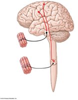
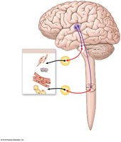
Divisions of the ANS
The ANS is divided into two main branches:
Sympathetic Division: Prepares the body for 'fight or flight' responses, increasing alertness, metabolic rate, and muscular abilities.
Parasympathetic Division: Promotes 'rest and digest' activities, conserving energy and maintaining resting metabolic rate.
These divisions often have opposing effects but may also work independently or cooperatively depending on the organ system involved.
Sympathetic Division
Organization and Pathways
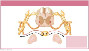
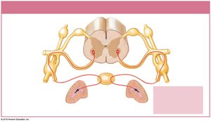
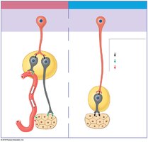
The sympathetic division is also known as the thoracolumbar division because its preganglionic neurons originate in the thoracic and lumbar regions (T1–L2) of the spinal cord. It is characterized by short preganglionic fibers and long postganglionic fibers. Sympathetic ganglia are located near the spinal cord and include:
Sympathetic chain ganglia: Located on either side of the vertebral column; innervate body wall, thoracic cavity, head, neck, and limbs.
Collateral ganglia: Located anterior to the vertebral bodies; innervate abdominopelvic organs.
Adrenal medullae: Modified sympathetic ganglia within the adrenal glands; release epinephrine and norepinephrine into the bloodstream.
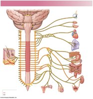
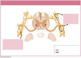
Sympathetic Neurotransmitters and Effects
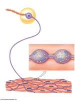
Sympathetic preganglionic neurons release acetylcholine (ACh) at ganglia, which is always excitatory. Most postganglionic neurons release norepinephrine (NE) (adrenergic), but some release ACh (cholinergic) or nitric oxide (NO) (nitroxidergic), depending on the target tissue. The effects of sympathetic activation include increased alertness, heart rate, blood pressure, respiratory rate, and mobilization of energy reserves.
Parasympathetic Division
Organization and Pathways
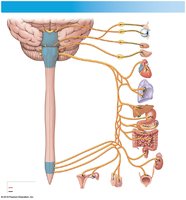
The parasympathetic division is also known as the craniosacral division because its preganglionic neurons originate in the brainstem and sacral spinal cord (S2–S4). It is characterized by long preganglionic fibers and short postganglionic fibers. Ganglia are located near or within target organs and are classified as:
Terminal ganglia: Located near target organs.
Intramural ganglia: Embedded within the tissues of target organs.
Major cranial nerves involved include III (oculomotor), VII (facial), IX (glossopharyngeal), and X (vagus). The vagus nerve provides the majority of parasympathetic outflow to thoracic and abdominopelvic organs.
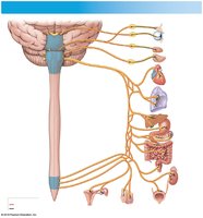
Parasympathetic Neurotransmitters and Effects
All parasympathetic neurons release acetylcholine (ACh). The effects depend on the type of cholinergic receptor present:
Nicotinic receptors: Always excitatory; found on ganglion cells of both divisions and at neuromuscular junctions.
Muscarinic receptors: Can be excitatory or inhibitory; found at neuroglandular junctions in the parasympathetic division.
Major effects include pupil constriction, increased digestive activity, stimulation of urination and defecation, and reduction in heart rate.
Comparison of Sympathetic and Parasympathetic Divisions
The sympathetic division has widespread effects due to extensive divergence and the release of hormones into the bloodstream, while the parasympathetic division has more specific, localized effects. The table below summarizes key differences:
Feature | Sympathetic Division | Parasympathetic Division |
|---|---|---|
Origin | Thoracolumbar (T1–L2) | Craniosacral (Brainstem, S2–S4) |
Preganglionic Fiber Length | Short | Long |
Postganglionic Fiber Length | Long | Short |
Main Neurotransmitter (Postganglionic) | Norepinephrine (NE) | Acetylcholine (ACh) |
General Effect | Widespread, long-lasting | Localized, short-lived |
Dual Innervation and Autonomic Tone
Most organs receive input from both sympathetic and parasympathetic divisions, often with opposing effects. Autonomic tone refers to the baseline level of activity in autonomic neurons, allowing for fine-tuned regulation. Some organs, such as blood vessels, are innervated by only one division (usually sympathetic).
Regulation of Autonomic Functions
Visceral Reflexes
Visceral reflexes are automatic, polysynaptic reflexes that regulate the activity of visceral effectors. They can be classified as:
Long reflexes: Involve the CNS and coordinate activities of entire organs.
Short reflexes: Bypass the CNS, involving only autonomic ganglia; control localized responses, especially in the digestive tract.
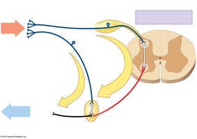
The enteric nervous system is a specialized network within the digestive tract capable of independent function.
Higher-Order Functions
Memory and Learning
Higher-order functions require the cerebral cortex and involve both conscious and unconscious processing. Memories are classified as:
Fact memories: Specific bits of information.
Skill memories: Learned motor behaviors.
Short-term memories: Easily recalled, but not retained long without repetition.
Long-term memories: More permanent; can be secondary (fade with time) or tertiary (permanent).
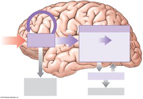
Key brain regions involved include the amygdaloid body, hippocampus, nucleus basalis, and cerebral cortex.
States of Consciousness and Sleep
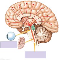
Consciousness is regulated by the reticular activating system (RAS) in the brainstem. Sleep consists of alternating periods of deep (NREM) and REM sleep, each with distinct physiological characteristics. The RAS is essential for arousal and maintaining wakefulness.
Effects of Aging on the Nervous System
Aging leads to anatomical and physiological changes in the nervous system, including reduced brain size, decreased number of neurons, reduced blood flow, and accumulation of abnormal proteins. These changes can impair memory, sensory perception, reflexes, and motor control. Most elderly individuals retain functional independence, but some develop senile dementia, with Alzheimer's disease being the most common form.
