 Back
The Autonomic Nervous System and Higher-Order Functions
Study Guide - Smart Notes
 Tailored notes based on your materials, expanded with key definitions, examples, and context.
Chapter 16: The Autonomic Nervous System and Higher-Order Functions
Introduction to the Autonomic Nervous System (ANS)
The autonomic nervous system (ANS), also known as the visceral motor system, is responsible for the unconscious regulation of vital physiological processes. It operates without conscious control and is essential for maintaining homeostasis by adjusting cardiovascular, respiratory, digestive, urinary, and reproductive functions. The ANS is divided into the sympathetic and parasympathetic divisions, each with distinct roles and mechanisms.
Comparison of the Somatic and Autonomic Nervous Systems
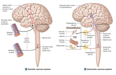
The somatic nervous system (SNS) and the autonomic nervous system (ANS) are both efferent divisions that carry motor commands, but they differ in their targets and organization:
SNS: Controls voluntary movements of skeletal muscles. Motor neurons in the CNS exert direct control over skeletal muscles.
ANS: Controls involuntary actions of visceral effectors such as smooth muscle, glands, cardiac muscle, and adipocytes. CNS motor neurons synapse on visceral motor neurons in autonomic ganglia, which then control visceral effectors.
Organization of the Autonomic Nervous System
The hypothalamus contains integrative centers for autonomic activity. The ANS consists of two-neuron chains:
Preganglionic neurons: Located in the brainstem and spinal cord; their axons (preganglionic fibers) synapse on ganglionic neurons in autonomic ganglia.
Ganglionic (postganglionic) neurons: Located in peripheral autonomic ganglia; their axons (postganglionic fibers) innervate visceral effectors.
Each autonomic ganglion acts as a relay station, where neurotransmitters are handed off from preganglionic to postganglionic fibers.
Divisions of the ANS
The ANS is divided into two main subdivisions:
Sympathetic Division: Prepares the body for emergencies ("fight or flight" response). Increases alertness, metabolic rate, heart rate, and blood pressure, while decreasing digestive and urinary functions.
Parasympathetic Division: Promotes relaxation and energy conservation ("rest and digest" response). Decreases heart rate and blood pressure, increases digestive activity, and stimulates urination and defecation.
Sympathetic Division of the ANS
Structural Organization
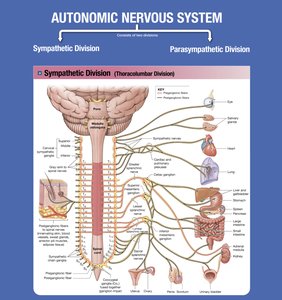
The sympathetic division is also known as the thoracolumbar division because its preganglionic neurons are located in the thoracic and lumbar segments (T1–L2) of the spinal cord. It is characterized by:
Short preganglionic fibers that synapse in ganglia near the spinal cord.
Long postganglionic fibers that extend to target organs.
There are three main pathways for sympathetic fibers:
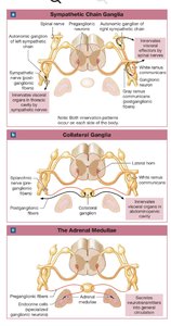
Sympathetic chain ganglia (paravertebral ganglia): Control effectors in the body wall, thoracic cavity, head, neck, and limbs.
Collateral ganglia (prevertebral ganglia): Innervate abdominopelvic tissues and viscera.
Adrenal medullae: Modified sympathetic ganglia that secrete epinephrine and norepinephrine into the bloodstream, acting as hormones.
Functional Organization and Effects
Sympathetic activation results in widespread physiological changes, including:
Increased alertness and energy
Elevated heart rate and blood pressure
Increased respiratory rate and dilation of airways
Decreased digestive and urinary activity
Activation of sweat glands
Neurotransmitters and Receptors in the Sympathetic Division
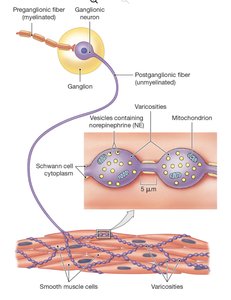
Sympathetic preganglionic neurons release acetylcholine (ACh) at synapses with ganglionic neurons (cholinergic synapses, always excitatory). Most sympathetic postganglionic neurons release norepinephrine (NE) at their varicosities (adrenergic synapses), but some release ACh (cholinergic) or nitric oxide (NO, nitroxidergic synapses).
Adrenergic receptors are divided into:
Alpha (α) receptors: α1 (excitatory, causes vasoconstriction), α2 (inhibitory, reduces cAMP levels)
Beta (β) receptors: β1 (increases metabolic activity and heart rate), β2 (inhibits smooth muscle, causes bronchodilation), β3 (stimulates lipolysis in adipose tissue)
Parasympathetic Division of the ANS
Structural Organization
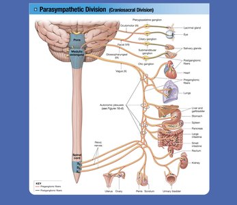
The parasympathetic division is also known as the craniosacral division because its preganglionic neurons are located in the brainstem and sacral segments (S2–S4) of the spinal cord. It is characterized by:
Long preganglionic fibers that synapse in ganglia near or within target organs (terminal or intramural ganglia).
Short postganglionic fibers that innervate target tissues.
Functional Organization and Effects
Parasympathetic activation is associated with energy conservation and restoration. Major effects include:
Constriction of pupils and focusing of the eyes
Stimulation of digestive gland secretion
Increased smooth muscle activity in the digestive tract
Stimulation of urination and defecation
Reduction in heart rate and force of contraction
Neurotransmitters and Receptors in the Parasympathetic Division
All parasympathetic neurons release acetylcholine (ACh). The effects depend on the type of cholinergic receptor present:
Nicotinic receptors: Found on ganglion cells of both divisions and at neuromuscular junctions; always excitatory.
Muscarinic receptors: Found at parasympathetic neuroeffector junctions; effects can be excitatory or inhibitory, depending on the target cell and second messenger systems.
Summary Table: Adrenergic and Cholinergic Receptors
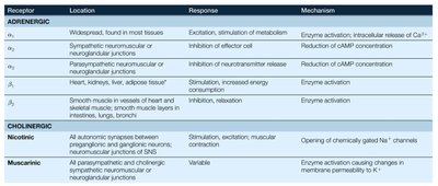
The following table summarizes the main types of receptors, their locations, responses, and mechanisms:
Receptor | Location | Response | Mechanism |
|---|---|---|---|
α1 (Adrenergic) | Widespread, found in most tissues | Excitation, stimulation of metabolism | Enzyme activation; intracellular release of Ca2+ |
α2 (Adrenergic) | Sympathetic neuromuscular or neuroglandular junctions; parasympathetic neuromuscular or neuroglandular junctions | Inhibition of effector cell; inhibition of neurotransmitter release | Reduction of cAMP concentration |
β1 (Adrenergic) | Heart, kidneys, liver, adipose tissue | Stimulation, increased energy consumption | Enzyme activation |
β2 (Adrenergic) | Smooth muscle in vessels of heart and skeletal muscle, smooth muscle layers in intestines, lungs, bronchi | Inhibition, relaxation | Enzyme activation |
β3 (Adrenergic) | Adipose tissue | Lipolysis | Enzyme activation |
Nicotinic (Cholinergic) | All autonomic synapses between preganglionic and ganglionic neurons; neuromuscular junctions of SNS | Stimulation, excitation, muscular contraction | Opening of chemically gated Na+ channels |
Muscarinic (Cholinergic) | All parasympathetic and cholinergic sympathetic neuromuscular or neuroglandular junctions | Variable | Enzyme activation causing changes in membrane permeability to K+ |
Clinical Application: Higher-Order Functions and Aging
Higher-order functions such as consciousness, learning, and intelligence are also influenced by the nervous system. Aging can affect memory, cognitive function, and the efficiency of the ANS, as illustrated by clinical cases of neurodegenerative diseases.
Key Equations and Concepts
Second Messenger Systems: Many ANS receptors are G protein–coupled, leading to the production of second messengers such as cyclic AMP (cAMP) or the release of intracellular Ca2+.
Neurotransmitter Inactivation: Norepinephrine is inactivated by monoamine oxidase (MAO) and catechol-O-methyltransferase (COMT); acetylcholine is inactivated by acetylcholinesterase (AChE).
Summary
The autonomic nervous system is essential for the regulation of involuntary physiological processes. Its sympathetic and parasympathetic divisions have distinct anatomical and functional characteristics, mediated by specific neurotransmitters and receptors. Understanding these mechanisms is crucial for comprehending how the body maintains homeostasis and responds to internal and external stimuli.
