 Back
BackThe Autonomic Nervous System and Homeostasis: Structure, Function, and Regulation
Study Guide - Smart Notes
 Tailored notes based on your materials, expanded with key definitions, examples, and context.
Chapter 14: The Autonomic Nervous System and Homeostasis
Overview of the Autonomic Nervous System (ANS)
The autonomic nervous system (ANS) is the involuntary branch of the peripheral nervous system (PNS), also known as the visceral motor division. It is responsible for regulating most vital functions, including heart rate, blood pressure, and digestive and urinary processes, without conscious control. The ANS is divided into two main divisions: the sympathetic and parasympathetic nervous systems, which work together to maintain homeostasis.
Functions of the ANS and Visceral Reflex Arcs
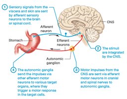
Visceral reflex arcs are the primary mechanism by which the ANS manages vital processes. Sensory stimuli from the viscera and skin are transmitted by afferent sensory neurons to the brain or spinal cord, where they are integrated by the CNS.
Motor impulses are then sent out via efferent neurons in cranial and spinal nerves to autonomic ganglia in the PNS, which relay signals to target organs, triggering a motor response.
This process exemplifies cell–cell communication in the nervous system.
Comparison of Somatic and Autonomic Nervous Systems
The motor division of the PNS is divided into the somatic and autonomic systems, each with distinct characteristics:
Somatic motor division: Neurons innervate skeletal muscle, producing voluntary muscle contractions initiated consciously.
Autonomic motor division: Neurons innervate smooth muscle, cardiac muscle, and glands, producing involuntary actions.
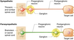
ANS motor neurons utilize a two-neuron circuit: a preganglionic neuron (cell body in CNS, releases acetylcholine) and a postganglionic neuron (cell body in autonomic ganglion in PNS, releases acetylcholine or norepinephrine).
Divisions of the Autonomic Nervous System
Sympathetic vs. Parasympathetic Nervous Systems
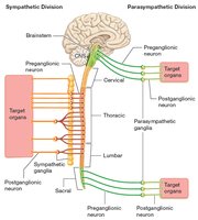
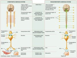
Sympathetic nervous system: Preganglionic axons are usually short; postganglionic axons are long. Preganglionic cell bodies originate in the thoracic and upper lumbar spinal cord (thoracolumbar division). Sympathetic ganglia are located near the spinal cord.
Parasympathetic nervous system: Preganglionic axons are long; postganglionic axons are short. Preganglionic cell bodies are located in the brainstem (cranial nerves) and sacral spinal cord (craniosacral division). Ganglia are near or within target organs.
Sympathetic Nervous System
Known as the "fight or flight" division, it prepares the body for emergency situations and physical activity.
It mediates responses to emotion and is vital for maintaining homeostasis during stress or exertion.
Parasympathetic Nervous System
Known as the "rest and digest" division, it is responsible for digestion, energy storage, and maintaining homeostasis at rest.
Cranial nerve X (vagus nerve) accounts for 75–90% of parasympathetic output, innervating thoracic and abdominal viscera.
Balance and Dual Innervation
The actions of the parasympathetic and sympathetic divisions are often antagonistic, maintaining a delicate balance for homeostasis.
Most organs receive dual innervation, allowing for precise regulation of physiological functions.
Study Boost: Pre- and Postganglionic Neurons
Ganglion: A cluster of neuronal cell bodies in the PNS.
Preganglionic neuron: The neuron before the ganglion; originates in the CNS.
Postganglionic neuron: The neuron after the ganglion; cell body in the ganglion, axon projects to the target organ.
Mnemonic: Think "first neuron" (preganglionic) and "second neuron" (postganglionic) to remember their order.
Module 14.2: The Sympathetic Nervous System
Gross and Microscopic Anatomy of the Sympathetic Nervous System
Sympathetic chain ganglia run parallel to the vertebral column, containing most postganglionic cell bodies.
Preganglionic neurons originate in the lateral horns of the thoracic and lumbar spinal cord, exit via the anterior root, and enter the sympathetic chain via white rami communicantes.
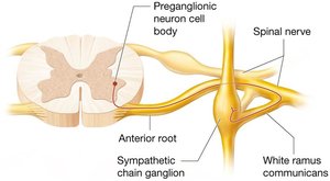
Some preganglionic axons pass through the chain ganglia to synapse in collateral ganglia near abdominopelvic organs (via splanchnic nerves).
Three possible synapse locations: (1) at the same level in the chain ganglion, (2) at a different level in the chain, or (3) in collateral ganglia.
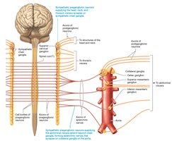
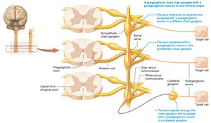
Postganglionic axons exit ganglia via gray rami communicantes to reach target cells.
Effects of the Sympathetic Nervous System on Target Cells
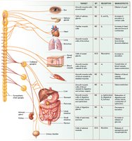
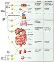
The sympathetic nervous system exerts widespread effects on various organs, preparing the body for rapid responses to stress or danger. These effects include increased heart rate, dilation of airways, inhibition of digestive activity, and mobilization of energy stores.
Module 14.3: The Parasympathetic Nervous System
Gross and Microscopic Anatomy of the Parasympathetic Nervous System
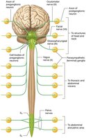
The parasympathetic division is known as the craniosacral division due to its association with cranial nerves and sacral spinal nerves.
Cranial nerves III (oculomotor), VII (facial), IX (glossopharyngeal), and X (vagus) carry parasympathetic fibers to various organs.
The vagus nerve is the primary parasympathetic nerve, innervating most thoracic and abdominal viscera.
Sacral nerves form pelvic splanchnic nerves, innervating the lower digestive tract, urinary bladder, and reproductive organs.
Parasympathetic Neurotransmitters and Receptors
Both pre- and postganglionic parasympathetic neurons release acetylcholine (ACh) at their synapses.
Two types of cholinergic receptors are present:
Nicotinic receptors: Found on all postganglionic neurons.
Muscarinic receptors: Found on all parasympathetic target cells.
Effects of the Parasympathetic Nervous System on Target Cells
The parasympathetic nervous system promotes activities that conserve and restore body energy, such as stimulating digestion, reducing heart rate, and promoting glandular secretion.
Module 14.4: Homeostasis Part II – PNS Maintenance of Homeostasis
Interactions of Autonomic Divisions
Both sympathetic and parasympathetic divisions innervate many of the same organs, where their actions are often antagonistic (dual innervation).
Sympathetic division dominates during physical activity, while the parasympathetic division maintains homeostasis during rest.
Autonomic Tone
Autonomic tone refers to the constant, baseline activity of both divisions of the ANS.
Sympathetic tone keeps blood vessels partially constricted, while parasympathetic tone maintains a resting heart rate of about 72 beats per minute.
Summary of Nervous System Control of Homeostasis
Homeostasis is centrally regulated by the hypothalamus and brainstem reticular formation, which coordinate the actions of the ANS.
Autonomic centers in the reticular formation contain neurons that control preganglionic sympathetic and parasympathetic neurons.
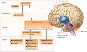
Table: Comparison of Sympathetic and Parasympathetic Nervous Systems
Feature | Sympathetic Nervous System | Parasympathetic Nervous System |
|---|---|---|
Main Role | "Fight or flight" – prepares body for emergencies, maintains homeostasis during activity | "Rest and digest" – conserves energy, maintains homeostasis at rest |
Origin of Preganglionic Neurons | Thoracic and lumbar regions of spinal cord | Brainstem (cranial nerves) and sacral spinal cord |
Location of Ganglia | Near spinal cord (sympathetic chain or collateral ganglia) | Near or within target organs (terminal ganglia) |
Neurotransmitters | Preganglionic: ACh; Postganglionic: ACh or norepinephrine | Preganglionic and postganglionic: ACh |
Receptors | Nicotinic (postganglionic), adrenergic (target cells) | Nicotinic (postganglionic), muscarinic (target cells) |
