 Back
BackThe Autonomic Nervous System and Homeostasis: Study Notes
Study Guide - Smart Notes
 Tailored notes based on your materials, expanded with key definitions, examples, and context.
The Autonomic Nervous System and Homeostasis
Overview of the Autonomic Nervous System (ANS)
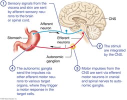
The Autonomic Nervous System (ANS) is a division of the peripheral nervous system that regulates involuntary physiological processes, including heart rate, blood pressure, digestion, and urinary functions. It operates largely without conscious control and is essential for maintaining homeostasis through visceral reflex arcs.
Visceral Reflex Arcs: Sensory signals from internal organs and skin are transmitted to the CNS, integrated, and then motor impulses are sent to target organs via autonomic ganglia.
Key Steps:
Sensory input from viscera/skin via afferent neurons to CNS
Integration in CNS
Motor output via efferent neurons to autonomic ganglia
Autonomic ganglia relay signals to target organs
Comparison of Somatic and Autonomic Nervous Systems
The Somatic Nervous System controls voluntary movements of skeletal muscles, while the Autonomic Nervous System controls involuntary actions of smooth muscle, cardiac muscle, and glands.
Somatic Motor Division: Single neuron pathway, neurotransmitter is acetylcholine (ACh), always excitatory.
Autonomic (Visceral) Motor Division: Two-neuron pathway (preganglionic and postganglionic), neurotransmitters include ACh and norepinephrine, can be excitatory or inhibitory.
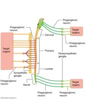
Divisions of the Autonomic Nervous System
Sympathetic and Parasympathetic Divisions
The ANS is divided into the Sympathetic and Parasympathetic divisions, which generally have opposing effects on target organs to maintain physiological balance.
Sympathetic Division: "Fight or Flight"; prepares the body for emergencies, increases heart rate, dilates pupils, inhibits digestion.
Parasympathetic Division: "Rest and Digest"; promotes maintenance activities, decreases heart rate, stimulates digestion, constricts pupils.
Anatomical Differences: Sympathetic preganglionic neurons originate in thoracic/lumbar spinal cord; parasympathetic in brainstem and sacral spinal cord.
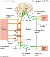
Balance and Dual Innervation
Most organs receive input from both divisions (dual innervation), allowing fine-tuned control. The two systems often act antagonistically to maintain homeostasis (e.g., sympathetic increases heart rate, parasympathetic decreases it).
Sympathetic Nervous System
Functions and Anatomy
The Sympathetic Nervous System prepares the body for physical activity and stress. It is also active during minor activities such as standing and emotional responses.
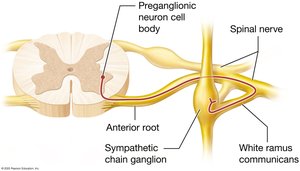
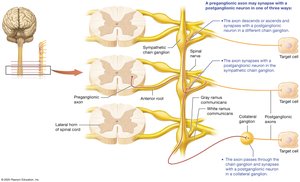
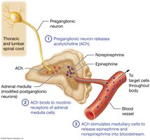
Preganglionic Neurons: Originate in the lateral horns of thoracic and lumbar spinal cord.
Sympathetic Chain Ganglia: Series of ganglia along the vertebral column; site of synapse for many pre- and postganglionic neurons.
White Rami Communicantes: Myelinated axons carrying preganglionic fibers to the ganglia.
Pathways and Ganglia
Sympathetic Chain Ganglia: Extend from cervical to sacral regions.
Collateral (Preaortic) Ganglia: Located near abdominal aorta; include celiac, superior mesenteric, and inferior mesenteric ganglia.
Splanchnic Nerves: Preganglionic fibers that pass through chain ganglia to synapse in collateral ganglia.
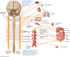
Three Pathways of Sympathetic Preganglionic Neurons
Synapse in the same chain ganglion
Ascend/descend to synapse in a different chain ganglion
Pass through chain ganglion to synapse in a collateral ganglion
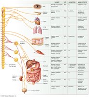
Neurotransmitters and Receptors
Sympathetic neurons use several neurotransmitters and receptors to mediate their effects:
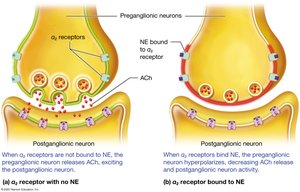
Preganglionic: Release acetylcholine (ACh) at synapse with postganglionic neuron (always excitatory).
Postganglionic: Most release norepinephrine (NE), some release epinephrine or ACh.
Adrenergic Receptors: Bind NE/epinephrine; include alpha (α1, α2) and beta (β1, β2, β3) subtypes, each with specific locations and effects.
Cholinergic Receptors: Bind ACh; include muscarinic (on sweat glands) and nicotinic (on all postganglionic neurons and adrenal medulla cells).
Effects on Target Cells
Sympathetic activation produces widespread physiological changes:
Cardiac Muscle: Increases heart rate and force of contraction.
Smooth Muscle: Vasoconstriction in digestive/urinary/skin vessels; vasodilation in skeletal/cardiac muscle; bronchodilation; pupil dilation; contraction of sphincters.
Metabolic Effects: Increases ATP production, stimulates breakdown of lipids and glycogen, increases blood glucose.
Adrenal Medulla: Releases epinephrine and norepinephrine into bloodstream, prolonging sympathetic effects.
Other Effects: Increases alertness, blood clotting, skeletal muscle tension, and triggers "goose bumps".
Pharmacology of the Sympathetic Nervous System
Antagonists (Blockers): Block adrenergic receptors, used to treat hypertension and other cardiovascular diseases.
Agonists: Mimic NE/epinephrine effects, used to treat asthma (bronchodilation) and other conditions.
Parasympathetic Nervous System
Functions and Anatomy
The Parasympathetic Nervous System is responsible for conserving energy and promoting maintenance activities during rest.
Preganglionic Neurons: Originate in brainstem (cranial nerves III, VII, IX, X) and sacral spinal cord (S2–S4).
Terminal Ganglia: Located near or within target organs; postganglionic axons are short.
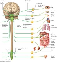
Parasympathetic Cranial and Sacral Nerves
Vagus Nerve (X): Provides ~90% of parasympathetic outflow, innervates heart, lungs, digestive tract.
Oculomotor (III), Facial (VII), Glossopharyngeal (IX): Innervate eyes, salivary glands, and other head structures.
Plexuses: Networks of nerves formed by branches of cranial and sacral nerves.
Neurotransmitters and Receptors
Both pre- and postganglionic neurons release ACh.
Nicotinic Receptors: On all postganglionic neurons (excitatory).
Muscarinic Receptors: On all target cells (excitatory or inhibitory depending on cell type).
Effects on Target Cells
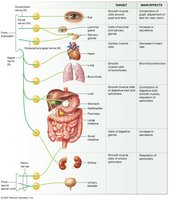
Cardiac Muscle: Decreases heart rate and blood pressure.
Smooth Muscle: Constricts pupils, accommodates lens for near vision, bronchoconstriction, stimulates peristalsis, relaxes sphincters, promotes urination and defecation, engorgement of erectile tissues.
Glandular Epithelial Cells: Stimulates secretion of tears, saliva, and digestive enzymes.
Metabolic Effects: No direct effect; promotes storage of energy reserves.
Pharmacology of the Parasympathetic Nervous System
Muscarinic Agonists: Stimulate parasympathetic effects; used to treat urinary retention and stimulate GI activity.
Muscarinic Antagonists: Block parasympathetic effects; used to treat bradycardia, motion sickness, and hypersalivation.
Anticholinergic Side Effects: Dry mouth, constipation, urinary retention, blurred vision.

Interactions and Regulation of the ANS
Dual Innervation and Autonomic Tone
Most organs receive input from both sympathetic and parasympathetic divisions, allowing precise regulation. Autonomic tone refers to the baseline level of activity in each system, with sympathetic tone dominating blood vessels and parasympathetic tone dominating the heart and digestive organs.
Homeostatic Control
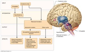
The hypothalamus and brainstem reticular formation are central regulators of autonomic function, integrating input from higher brain centers and controlling preganglionic neurons. Emotional states can influence ANS activity via the amygdala and cerebral cortex.
Clinical Correlation: Postural Orthostatic Tachycardia Syndrome (POTS)
POTS is characterized by an abnormal increase in heart rate upon standing, often accompanied by dizziness and other symptoms. It is thought to involve excessive sympathetic activity or sensitivity to catecholamines. Treatment includes lifestyle modifications and medications to block sympathetic receptors.
Summary Table: Comparison of Sympathetic and Parasympathetic Divisions
Feature | Sympathetic Division | Parasympathetic Division |
|---|---|---|
Origin | Thoracic & lumbar spinal cord | Brainstem & sacral spinal cord |
Ganglia Location | Near spinal cord (chain/collateral) | Near or within target organs |
Neurotransmitters | ACh (preganglionic), NE/Epi (postganglionic) | ACh (both pre- and postganglionic) |
Main Functions | "Fight or Flight" (emergency, stress) | "Rest and Digest" (maintenance, rest) |
Effects on Heart | Increases rate & force | Decreases rate |
Effects on Digestive System | Inhibits activity | Stimulates activity |
Pupil | Dilates | Constricts |
