Back
BackThe Autonomic Nervous System and Visceral Reflexes: Structure, Function, and Control CH 15
Study Guide - Smart Notes
 Tailored notes based on your materials, expanded with key definitions, examples, and context.
The Autonomic Nervous System (ANS)
General Properties of the ANS
The autonomic nervous system (ANS), also known as the visceral motor system, is a motor nervous system that controls glands, cardiac muscle, and smooth muscle. It operates largely without conscious control and regulates the function of internal organs to maintain homeostasis.
Primary target organs: Viscera of thoracic and abdominal cavities, cutaneous blood vessels, sweat glands, and arrector muscles.
Involuntary action: The ANS adjusts organ activity to the body's needs, not to initiate function.
Denervation hypersensitivity: Organs can show exaggerated responses if ANS nerves are severed.
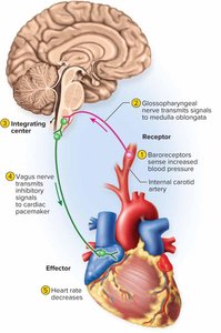
Visceral Reflexes
Visceral reflexes are unconscious, automatic, and stereotyped responses to stimulation involving visceral receptors and effectors, controlled by the ANS. The reflex arc includes:
Receptors: Detect stretch, tissue damage, blood chemicals, temperature, and other internal stimuli.
Afferent neurons: Carry signals to the CNS.
Integrating center: Interneurons in the CNS process the information.
Efferent neurons: Carry motor signals away from the CNS.
Effectors: Carry out the end response.
Divisions of the ANS
Sympathetic and Parasympathetic Divisions
The ANS has two main divisions with generally opposing effects:
Sympathetic division: Prepares the body for physical activity (fight-or-flight response). Increases heart rate, blood pressure, airflow, and blood glucose; reduces blood flow to skin and digestive tract.
Parasympathetic division: Calms body functions, reduces energy expenditure, and assists in bodily maintenance (rest-and-digest state). Promotes digestion and waste elimination.
Autonomic tone is the background rate of activity representing the balance between the two divisions, adjusting according to the body's needs.
Anatomy of the Autonomic Nervous System
Autonomic Output Pathways
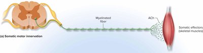
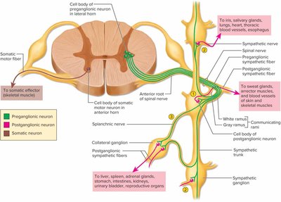
The ANS has components in both the central and peripheral nervous systems. Its motor pathway differs from the somatic motor pathway:
Somatic pathway: A single myelinated motor neuron extends from the CNS to the skeletal muscle.
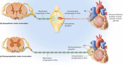
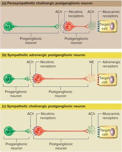
Autonomic pathway: Involves two neurons and a synapse in an autonomic ganglion:
Preganglionic neuron: Cell body in CNS; axon (preganglionic fiber) extends to autonomic ganglion.
Postganglionic neuron: Cell body in ganglion; axon (postganglionic fiber) extends to target cells.
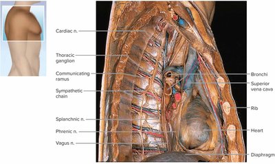
The Sympathetic Division
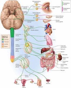
The sympathetic division (thoracolumbar division) arises from the thoracic and lumbar regions of the spinal cord. It has short preganglionic and long postganglionic fibers. Preganglionic neurons originate in the lateral horns of the spinal cord (T1–L2) and synapse in the sympathetic chain (paravertebral) ganglia.
Sympathetic fibers are distributed throughout the body.
Each ganglion is connected to a spinal nerve by communicating rami (white and gray).
Nerve fibers leave the sympathetic chain by three routes: spinal, sympathetic, and splanchnic nerves.

Collateral Ganglia and Abdominal Plexuses
Collateral ganglia form the abdominal aortic plexus, which includes the celiac, superior mesenteric, and inferior mesenteric ganglia. These ganglia distribute sympathetic fibers to abdominal organs.
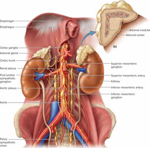
The Adrenal Glands
The adrenal (suprarenal) glands are located on the superior poles of the kidneys. Each gland has:
Adrenal cortex: Secretes steroid hormones.
Adrenal medulla: Functions as a sympathetic ganglion, releasing catecholamines (epinephrine and norepinephrine) into the bloodstream.
The sympathoadrenal system refers to the close relationship between the adrenal medulla and the sympathetic nervous system.
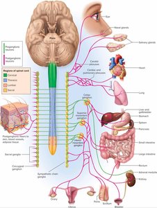
The Parasympathetic Division
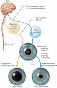
The parasympathetic division (craniosacral division) arises from the brain and sacral spinal cord. It has long preganglionic and short postganglionic fibers. Preganglionic neurons originate in the brainstem (cranial nerves III, VII, IX, X) and sacral spinal cord (S2–S4).
Parasympathetic fibers are relatively selective in their effects due to little neural divergence.
Pelvic splanchnic nerves supply the distal colon, rectum, bladder, and reproductive organs.
The Enteric Plexus
The enteric nervous system is a network of neurons in the digestive tract wall, regulating motility and secretion. It operates independently but is influenced by the sympathetic and parasympathetic divisions.
Hirschsprung disease: Absence of enteric plexus in part of the colon, leading to megacolon and severe constipation.
Autonomic Effects on Target Organs
Neurotransmitters and Their Receptors
The sympathetic and parasympathetic divisions use different neurotransmitters and receptors, allowing for diverse effects on target organs.
Acetylcholine (ACh): Secreted by all preganglionic neurons and parasympathetic postganglionic neurons. Receptors are cholinergic (muscarinic and nicotinic).
Norepinephrine (NE): Secreted by most sympathetic postganglionic neurons. Receptors are adrenergic (alpha and beta).
Other neurotransmitters: Enkephalin, substance P, neuropeptide Y, nitric oxide, etc.
Dual Innervation
Most organs receive both sympathetic and parasympathetic innervation, which may have antagonistic (opposing) or cooperative effects. For example, the heart rate is slowed by parasympathetic and increased by sympathetic stimulation. In the iris, each division innervates different muscle groups to control pupil size.
Control Without Dual Innervation
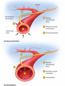
Some effectors, such as blood vessels, sweat glands, and the adrenal medulla, receive only sympathetic innervation. Sympathetic tone maintains vasomotor tone, regulating blood pressure and blood flow distribution.
Central Control of Autonomic Function
Brain and Spinal Cord Regulation
The ANS is regulated at multiple CNS levels:
Cerebral cortex and limbic system: Emotional states influence ANS activity.
Hypothalamus: Major control center for visceral motor functions (hunger, thirst, thermoregulation, etc.).
Midbrain, pons, medulla oblongata: Contain nuclei for cardiac, vasomotor, salivary, and digestive control.
Spinal cord: Integrates autonomic reflexes for defecation and urination.
Drugs and the Autonomic Nervous System
Neuropharmacology
Drugs can enhance or suppress autonomic activity:
Sympathomimetics: Enhance sympathetic activity (e.g., cold medicines).
Sympatholytics: Suppress sympathetic activity (e.g., beta-blockers).
Parasympathomimetics: Enhance parasympathetic effects.
Parasympatholytics: Suppress parasympathetic effects.
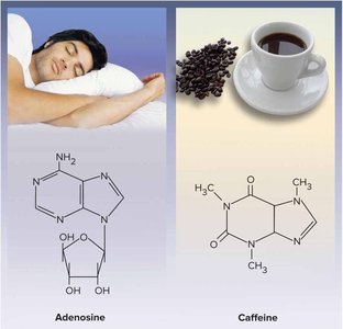
Other drugs act on CNS neurotransmitters (e.g., SSRIs, MAO inhibitors, caffeine).