 Back
BackThe Autonomic Nervous System (ANS): Structure, Function, and Neurotransmission
Study Guide - Smart Notes
 Tailored notes based on your materials, expanded with key definitions, examples, and context.
Introduction to the Autonomic Nervous System (ANS)
Overview of the Peripheral Nervous System
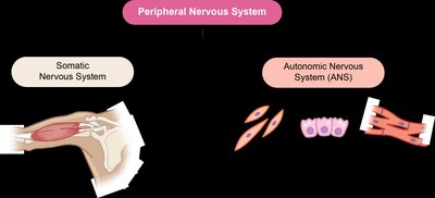
The peripheral nervous system (PNS) is divided into the somatic nervous system and the autonomic nervous system (ANS). The somatic system controls voluntary movements of skeletal muscles, while the ANS regulates involuntary functions by innervating smooth muscle, cardiac muscle, and glands.
Somatic Nervous System: Controls voluntary skeletal muscle movement; single-neuron efferent pathway.
Autonomic Nervous System: Controls involuntary actions in smooth muscle, cardiac muscle, and glands; two-neuron efferent pathway (preganglionic and postganglionic neurons).
Divisions of the Autonomic Nervous System
Sympathetic and Parasympathetic Divisions
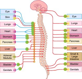
The ANS is divided into the sympathetic and parasympathetic divisions, which typically have opposing effects on target organs to maintain homeostasis.
Sympathetic Division: Activated during stress, threat, or physical activity ("fight or flight").
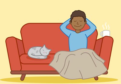
Parasympathetic Division: Dominates during rest and relaxation ("rest and digest").
Most organs receive dual innervation from both divisions, which work antagonistically.

Control of the Autonomic Nervous System
Relevant Brain Anatomy
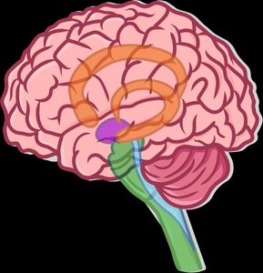
Several brain regions regulate the ANS, integrating sensory input and coordinating autonomic output.
Hypothalamus: The main integration center for autonomic functions (e.g., heart rate, blood pressure, digestion).
Brainstem: Relays information between the brain and body; contains nuclei for autonomic control.
Reticular Formation: Regulates skeletal and visceral muscle activity; influences autonomic functions.
Limbic System: Processes emotional stimuli and can influence autonomic responses.
Levels of Control in the ANS
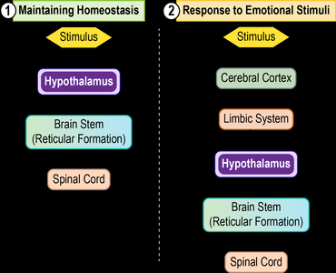
The hypothalamus integrates signals from higher brain centers and coordinates autonomic responses. The ANS operates to maintain homeostasis and respond to emotional or stressful stimuli via distinct neural pathways.
Homeostatic Regulation: Hypothalamus → Brainstem (Reticular Formation) → Spinal Cord
Emotional Response: Cerebral Cortex → Limbic System → Hypothalamus → Brainstem → Spinal Cord
Sympathetic Nervous System
Function of the Sympathetic Nervous System
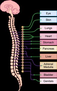
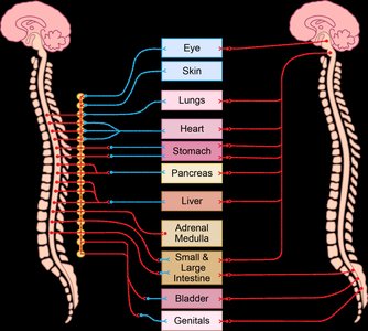
The sympathetic division prepares the body for physical activity or stressful situations by increasing heart rate, dilating bronchioles, mobilizing energy stores, and inhibiting non-essential functions like digestion.
Cardiac Muscle: Increases heart rate
Bronchioles: Dilates airways
Liver: Releases glucose
Gastrointestinal Tract: Decreases activity
Blood Vessels: Vasoconstriction in viscera, vasodilation in skeletal muscle
Pupils: Dilate
Sweat Glands: Stimulated
Structure of the Sympathetic Nervous System
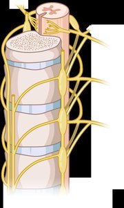
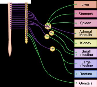
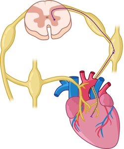
Sympathetic fibers originate from the thoracolumbar region of the spinal cord. The sympathetic trunk (chain of ganglia) runs alongside the vertebral column. Preganglionic fibers are generally short, and postganglionic fibers are long.
White Ramus Communicans: Carries preganglionic fibers to sympathetic ganglia.
Gray Ramus Communicans: Carries postganglionic fibers to spinal nerves.
Pathways of Sympathetic Innervation
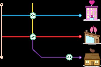
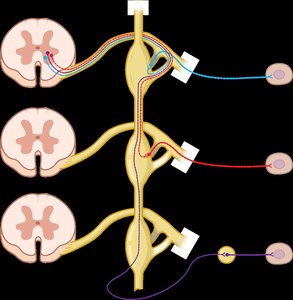
Preganglionic neurons can synapse with postganglionic neurons in the sympathetic trunk at the same level, a higher/lower level, or in collateral ganglia. All impulses must synapse once before reaching effectors.
Splanchnic Nerves and the Adrenal Medulla
Splanchnic nerves bypass the sympathetic trunk and synapse in collateral ganglia, innervating abdominal organs. The adrenal medulla acts as a modified sympathetic ganglion, releasing epinephrine and norepinephrine into the bloodstream.
Parasympathetic Nervous System
Function of the Parasympathetic Nervous System
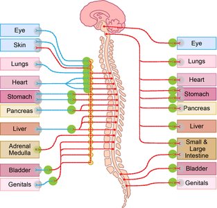
The parasympathetic division promotes rest, energy conservation, and restoration. It slows the heart rate, increases digestive activity, and facilitates urination and defecation.
Cardiac Muscle: Decreases heart rate
Bronchioles: Constricts airways
Gastrointestinal Tract: Increases activity
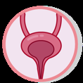
Urinary Sphincters: Relaxes smooth muscle for urination
Pupils: Constrict

Structure of the Parasympathetic Nervous System
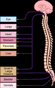
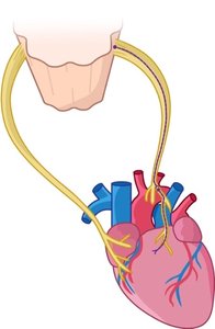
Parasympathetic fibers arise from the brainstem and sacral spinal cord (craniosacral division). The vagus nerve (CN X) provides most parasympathetic innervation. Preganglionic fibers are long and synapse in terminal ganglia near or within target organs; postganglionic fibers are short.
Review: Dual Innervation of the Heart
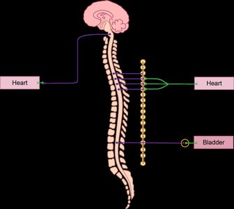
Both sympathetic and parasympathetic divisions innervate the heart, but with opposite effects. The sympathetic division increases heart rate, while the parasympathetic division decreases it. Sympathetic fibers synapse in the trunk ganglia; parasympathetic fibers synapse in terminal ganglia.
Visceral Reflex Arcs
Structure and Function
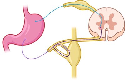
Visceral reflex arcs control involuntary responses in smooth muscle, cardiac muscle, and glands. They involve two consecutive motor neurons (preganglionic and postganglionic), visceral sensory neurons, and an integration center (often in the CNS).
Five steps: Receptor → Visceral sensory neuron → Integration center → Motor neuron (2-neuron chain) → Visceral effector
Neurotransmitters of the ANS
Major Neurotransmitters
The ANS uses two main neurotransmitters: acetylcholine (ACh) and norepinephrine (NE). Their release and effects depend on the division and location within the pathway.
Division | Preganglionic Fibers Release | Postganglionic Fibers Release |
|---|---|---|
Sympathetic | Acetylcholine (ACh) | Mostly Norepinephrine (NE), some Epinephrine (E) |
Parasympathetic | Acetylcholine (ACh) | Acetylcholine (ACh) |
Cholinergic fibers: Release ACh (all preganglionic fibers, all parasympathetic postganglionic fibers).
Adrenergic fibers: Release NE (most sympathetic postganglionic fibers).
Neurotransmitter Receptors
Receptors are classified by the neurotransmitter they bind:
Cholinergic Receptors: Bind ACh; include nicotinic (at all ganglia and adrenal medulla) and muscarinic (at parasympathetic target organs and some sympathetic targets like sweat glands).
Adrenergic Receptors: Bind NE and E; include alpha (α) and beta (β) subtypes, found at most sympathetic target organs.
Summary Table: Comparison of Somatic and Autonomic Nervous Systems
Feature | Somatic Nervous System | Autonomic Nervous System |
|---|---|---|
Effector Organs | Skeletal muscle | Smooth muscle, cardiac muscle, glands |
Control | Voluntary | Involuntary |
Number of Neurons | One | Two (pre- and postganglionic) |
Neurotransmitter | Acetylcholine (ACh) | ACh, Norepinephrine (NE), Epinephrine (E) |
Effect | Always excitatory | Excitatory or inhibitory |
