 Back
BackThe Autonomic Nervous System (ANS): Structure, Function, and Divisions
Study Guide - Smart Notes
 Tailored notes based on your materials, expanded with key definitions, examples, and context.
The Autonomic Nervous System (ANS)
Overview of the ANS
The Autonomic Nervous System (ANS) is a critical component of the peripheral nervous system responsible for involuntary control of body functions. It regulates the activity of smooth muscle, cardiac muscle, and glands, maintaining homeostasis without conscious effort.
Effectors: The ANS innervates cardiac muscle, smooth muscle, and glands, while the somatic nervous system innervates skeletal muscle.
Control: Operates under subconscious control, continuously adjusting internal conditions.
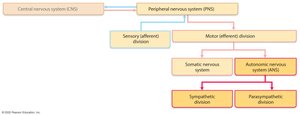
Structural Organization of the Nervous System
The nervous system is divided into the central nervous system (CNS) and peripheral nervous system (PNS). The PNS is further divided into sensory (afferent) and motor (efferent) divisions. The motor division includes the somatic and autonomic nervous systems. The ANS is subdivided into the sympathetic and parasympathetic divisions.
Comparison: Somatic vs. Autonomic Nervous Systems
Effectors
Somatic Nervous System: Controls voluntary movements by innervating skeletal muscles.
Autonomic Nervous System: Controls involuntary responses by innervating cardiac muscle, smooth muscle, and glands.
Efferent Pathways and Ganglia
Somatic Pathway: Single, thick, myelinated axon extends from the CNS directly to skeletal muscle; no motor ganglia involved.
Autonomic Pathway: Utilizes a two-neuron chain:
Preganglionic neuron: Cell body in CNS; thin, lightly myelinated axon extends to autonomic ganglion.
Postganglionic neuron: Cell body in autonomic ganglion outside CNS; nonmyelinated axon extends to effector organ.
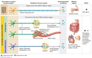
Neurotransmitter Effects
Somatic Nervous System: All motor neurons release acetylcholine (ACh); effect is always stimulatory.
Autonomic Nervous System: Preganglionic fibers release ACh; postganglionic fibers release norepinephrine (sympathetic) or ACh (parasympathetic). Effects can be stimulatory or inhibitory, depending on the receptor type.
Divisions of the Autonomic Nervous System
Sympathetic and Parasympathetic Divisions
The ANS is divided into two main branches that typically have opposing effects on target organs:
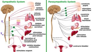
Parasympathetic Division: Promotes maintenance functions and conserves energy; known as the "rest and digest" system.
Sympathetic Division: Mobilizes the body during activity; known as the "fight or flight" system.
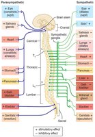
Dual Innervation and Homeostasis
Most visceral organs receive input from both divisions, which typically exert opposite effects to maintain homeostasis.
Role of the Parasympathetic Division
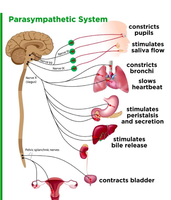
The parasympathetic division is active during restful states, promoting energy conservation and routine maintenance activities.
Decreases blood pressure, heart rate, and respiratory rate.
Increases gastrointestinal activity after meals.
Constricts pupils.
Role of the Sympathetic Division
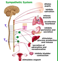
The sympathetic division prepares the body for stressful or energetic activity.
Increases heart rate and dilates pupils.
Shunts blood to skeletal muscles and heart, away from the skin.
Dilates bronchioles and stimulates glucose release from the liver.
Anatomical Differences Between Divisions
Sites of Origin
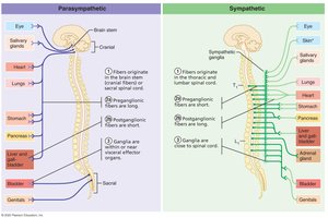
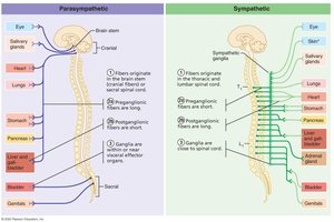
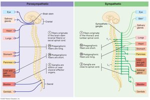
Parasympathetic fibers: Originate from the brainstem and sacral spinal cord (craniosacral).
Sympathetic fibers: Originate from the thoracic and lumbar regions of the spinal cord (thoracolumbar).
Fiber Lengths
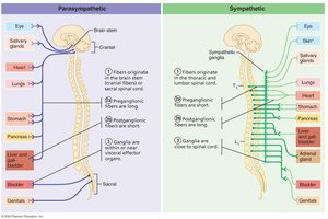
Parasympathetic: Long preganglionic and short postganglionic fibers.
Sympathetic: Short preganglionic and long postganglionic fibers.
Location of Ganglia
Parasympathetic: Ganglia are located in or near the effector organs.
Sympathetic: Ganglia are close to the spinal cord.
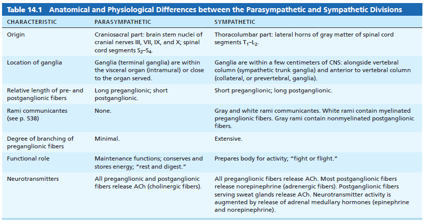
Summary Table: Anatomical and Physiological Differences
Characteristic | Parasympathetic | Sympathetic |
|---|---|---|
Origin | Craniosacral part: brain stem nuclei of cranial nerves III, VII, IX, X; spinal cord segments S2–S4 | Thoracolumbar part: lateral horns of gray matter of spinal cord segments T1–L2 |
Location of ganglia | Within or near visceral organ | Close to CNS: alongside or anterior to vertebral column |
Relative length of fibers | Long preganglionic; short postganglionic | Short preganglionic; long postganglionic |
Rami communicantes | None | Gray and white rami communicantes |
Degree of branching | Minimal | Extensive |
Functional role | Maintenance, "rest and digest" | "Fight or flight" |
Neurotransmitters | All fibers release ACh | Preganglionic: ACh; Postganglionic: NE (except sweat glands: ACh) |
Parasympathetic Division Pathways
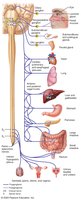
Cranial Part
Vagus nerve (X): Accounts for ~90% of all preganglionic fibers; serves thoracic and abdominal viscera.
Fibers arise from the medulla and synapse in terminal ganglia within target organs.
Cardiac, pulmonary, and esophageal plexuses regulate heart, lungs, and digestive organs.
Sacral Part
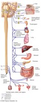
Originates from S2–S4 spinal segments; serves pelvic organs and distal large intestine.
Axons travel in ventral roots, forming pelvic splanchnic nerves to synapse in terminal ganglia of target organs.
Sympathetic Division Pathways
General Organization
Preganglionic cell bodies are in the lateral horns of the spinal cord (T1–L2).
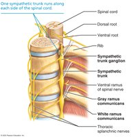
Axons exit via ventral roots, pass through white rami communicantes, and enter sympathetic trunk ganglia.
Sympathetic division innervates more organs than the parasympathetic division, including structures unique to it (e.g., sweat glands, arrector pili muscles).
Three Pathways Upon Entering the Sympathetic Trunk
Synapse in the same trunk ganglion: Typically for effectors in the skin.
Ascend or descend to another trunk ganglion: For effectors such as blood vessels.
Pass through trunk without synapsing: Synapse in collateral ganglia (abdomen/pelvis), forming splanchnic nerves.
Visceral Reflexes
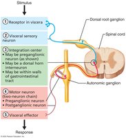
Visceral reflexes are automatic responses involving the ANS, controlling functions such as digestion, heart rate, and respiratory rate. These reflexes involve receptors, sensory neurons, integration centers, motor neurons, and visceral effectors.
Control of ANS Function
The ANS is regulated by the CNS, primarily through the hypothalamus, brainstem, and spinal cord. The hypothalamus serves as the main integrative center, while the cerebral cortex can influence ANS activity subconsciously via the limbic system.
Hypothalamus: Main control center for ANS activity.
Brainstem and spinal cord: Mediate basic ANS reflexes.
Cerebral cortex: Modifies ANS activity through emotional and subconscious pathways.
