 Back
BackThe Autonomic Nervous System: Structure, Function, and Neurotransmission
Study Guide - Smart Notes
 Tailored notes based on your materials, expanded with key definitions, examples, and context.
Autonomic Nervous System (ANS)
Overview and General Organization
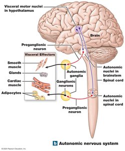
The Autonomic Nervous System (ANS) is a division of the peripheral nervous system responsible for involuntary regulation of internal organs and homeostatic processes. It controls visceral effectors such as smooth muscle, cardiac muscle, glands, and adipose tissue. The ANS operates through a two-neuron pathway: a preganglionic neuron originating in the CNS and a postganglionic neuron in the peripheral ganglia, which innervates the target organ.
Visceral motor nuclei in the hypothalamus initiate autonomic responses.
Preganglionic neurons originate in the brainstem or spinal cord.
Autonomic ganglia house the cell bodies of postganglionic neurons.
Postganglionic axons extend to visceral effectors.
Divisions of the ANS
The ANS is divided into two main branches with generally opposing effects:
Sympathetic Division – Prepares the body for emergency situations ("fight or flight").
Parasympathetic Division – Promotes maintenance activities during restful states ("rest and digest").
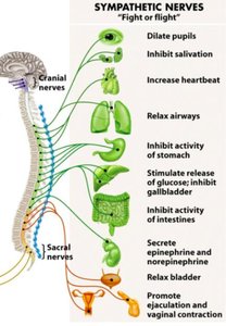
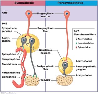
Sympathetic Division
Structure and Pathways
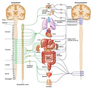
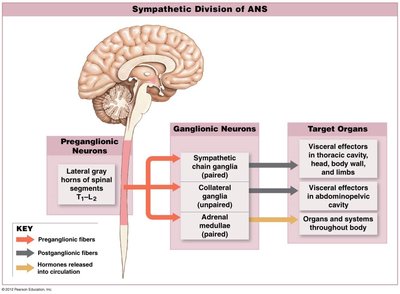
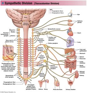
The sympathetic division is organized to mobilize the body's resources during stress or emergencies. Preganglionic fibers originate in the thoracic and lumbar segments (T1–L2) of the spinal cord. There are three main types of sympathetic ganglia:
Chain ganglia – Located alongside the spinal cord; control effectors in the body wall, thoracic cavity, head, neck, and limbs.
Collateral ganglia – Anterior to the spinal cord; control effectors in abdominopelvic tissues and organs.
Adrenal medullae – Modified ganglia within the adrenal glands; release hormones directly into the bloodstream.
Key Characteristics
Short preganglionic axons (originate from T1–L2).
Ganglia are close to the spinal cord.
Long postganglionic axons.
One preganglionic neuron may innervate many targets.
Functions of the Sympathetic Division
Sympathetic activation produces a coordinated set of physiological changes:
Heightened mental alertness
Pupil dilation
Increased metabolic rate and ATP production
Reduced digestive and urinary functions
Activation of energy reserves (glycogenolysis, lipolysis)
Increased respiratory rate and dilation of airways
Increased heart rate and blood pressure
Activation of sweat glands
Orgasm (sexual function)
Activation and Systemic Effects
Sympathetic responses often involve the entire division activating simultaneously (mass activation).
Results in increased alertness, energy, euphoria, and temporary insensitivity to pain.
Mobilization of energy reserves (glycogen breakdown, lipid release).
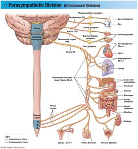
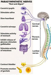
Parasympathetic Division
Structure and Pathways
The parasympathetic division is responsible for conserving energy and promoting maintenance activities. Preganglionic fibers originate in the brainstem (cranial nerves III, VII, IX, X) and sacral spinal cord segments (S2–S4). Ganglia are located near or within target organs:
Terminal ganglia – Near target organ
Intramural ganglia – Embedded within the target organ's tissue
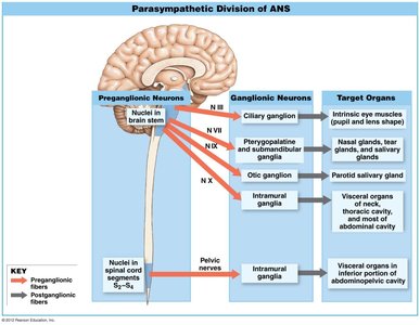
Key Characteristics
Long preganglionic axons (from brainstem and sacral region)
Ganglia within or adjacent to target organs
Short postganglionic axons
One neuron innervates specific targets (more localized effects)
Functions of the Parasympathetic Division
Parasympathetic activation supports rest, digestion, and energy storage:
Decreased metabolic rate and ATP production
Decreased heart rate and blood pressure
Constriction of respiratory passages
Increased secretion by salivary and digestive glands
Increased blood flow to digestive tract
Increased absorption of nutrients
Stimulation of urination and defecation
Constriction of pupils
Sexual arousal and erection
Activation and Systemic Effects
Parasympathetic functions are the default state and are not activated as a single unit.
Focuses on relaxation, food processing, and nutrient absorption.
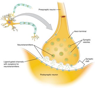
Neurotransmitters and Receptors in the ANS
Acetylcholine (ACh)
Released by cholinergic neurons (all preganglionic neurons and parasympathetic postganglionic neurons).
Effects are brief due to rapid breakdown by acetylcholinesterase (AChE).
Nicotinic receptors – Found in autonomic ganglia and neuromuscular junctions; always excitatory (ionotropic).
Muscarinic receptors – Found on parasympathetic target organs; can be excitatory or inhibitory (metabotropic, G-protein coupled).
Norepinephrine (NE) and Epinephrine
Released by adrenergic neurons (most sympathetic postganglionic neurons).
Effects last longer than ACh; NE is reabsorbed and degraded by monoamine oxidase (MAO) and catechol-O-methyltransferase (COMT).
Alpha (\( \alpha \)) receptors – \( \alpha_1 \) (excitatory), \( \alpha_2 \) (inhibitory, often inhibits parasympathetic activity).
Beta (\( \beta \)) receptors – \( \beta_1 \) (increases heart rate/metabolism), \( \beta_2 \) (relaxes smooth muscle in airways), \( \beta_3 \) (stimulates lipolysis in adipose tissue).
Summary Table: Neurotransmitters and Receptors
Division | Preganglionic Neurotransmitter (Receptor) | Postganglionic Neurotransmitter (Receptor) |
|---|---|---|
Sympathetic | Acetylcholine (Nicotinic) | Mostly Norepinephrine (Alpha and Beta) |
Parasympathetic | Acetylcholine (Nicotinic) | Acetylcholine (Muscarinic) |
Comparison of Sympathetic and Parasympathetic Divisions
Characteristic | Sympathetic | Parasympathetic |
|---|---|---|
Origin of Preganglionic Neuron | Spinal Segments T1–L2 | Cranial Nerves III, VII, IX, X; Sacral Segments S2–S4 |
Location of Peripheral Ganglia | Near spinal cord | Near or within target organ/tissue |
Preganglionic Fiber Length | Short | Long |
Postganglionic Fiber Length | Long | Short |
Preganglionic Neurotransmitter (receptor) | ACh (Nicotinic) | ACh (Nicotinic) |
Postganglionic Neurotransmitter (receptor) | Mostly Norepinephrine (Alpha and Beta) | Acetylcholine (Muscarinic) |
Target Area | Broad | Specific |
General Function | Stimulates metabolism, increases alertness, prepares for emergency | Promotes relaxation, nutrient uptake, energy storage |
