Back
BackThe Autonomic Nervous System: Structure, Function, and Integration
Study Guide - Smart Notes
 Tailored notes based on your materials, expanded with key definitions, examples, and context.
Autonomic Nervous System Overview
Introduction to the Autonomic Nervous System (ANS)
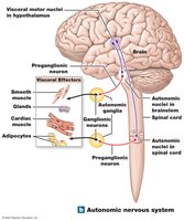
The autonomic nervous system (ANS) is a division of the peripheral nervous system responsible for involuntary control of visceral effectors, including smooth muscle, glands, cardiac muscle, and adipocytes. It coordinates essential functions such as cardiovascular, respiratory, digestive, urinary, and reproductive activities. The integrative centers of the ANS are located in the hypothalamus, and its motor neurons synapse in autonomic ganglia outside the central nervous system (CNS).
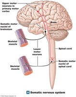
Comparison: Autonomic vs. Somatic Nervous System
Somatic Nervous System (SNS): Controls voluntary movements of skeletal muscles. CNS motor neurons synapse directly with skeletal muscle cells.
Autonomic Nervous System (ANS): Controls involuntary actions of visceral organs. CNS visceral motor neurons synapse with visceral motor neurons in autonomic ganglia, which then innervate target organs.
Organization of the Autonomic Nervous System
Visceral Motor Neurons
Preganglionic neurons: Cell bodies in the brainstem and spinal cord; their axons (preganglionic fibers) project to autonomic ganglia.
Postganglionic neurons: Cell bodies in autonomic ganglia; their axons (postganglionic fibers) innervate peripheral target organs.
Divisions of the ANS
Sympathetic Division: Prepares the body for stress or emergency ("fight or flight"). Increases alertness, respiratory rate, metabolic rate, and muscular abilities.
Parasympathetic Division: Conserves energy and maintains resting metabolic rate ("rest and digest").
Functional Interactions
Sympathetic and parasympathetic divisions usually have opposing effects (excitation vs. inhibition).
Some structures are innervated by only one division, or both may work together in complex processes.
The Sympathetic Division
Organization and Pathways
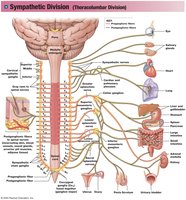
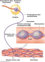
The sympathetic division is also known as the thoracolumbar division, with preganglionic neuron cell bodies located in the lateral horns of the thoracic and lumbar spinal cord (T1–L2). Preganglionic fibers are short and synapse with many postganglionic neurons, releasing acetylcholine (ACh). Postganglionic fibers are long and release norepinephrine (NE) at target organs.
Types of Sympathetic Ganglia
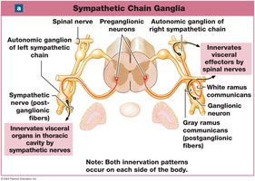
Sympathetic chain ganglia (paravertebral ganglia): Located on either side of the vertebral column; innervate effectors in the body wall, thoracic cavity, head, neck, and limbs.
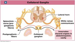
Collateral ganglia (prevertebral ganglia): Anterior to the vertebral column; innervate abdominopelvic tissues and viscera.
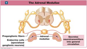
Adrenal medullae: Modified sympathetic ganglia within the adrenal glands; release epinephrine and norepinephrine into the bloodstream.
Effects of Sympathetic Activation
Heightened mental alertness
Increased metabolic rate
Reduced digestive and urinary functions
Activation of energy reserves
Increased respiratory rate and dilation of passageways
Increased heart rate and blood pressure
Activation of sweat glands
Sympathetic Neurotransmitters and Receptors
Neurotransmitters
Preganglionic neurons release acetylcholine (ACh) at ganglia (cholinergic synapses; always excitatory).
Most postganglionic neurons release norepinephrine (NE) at target organs (adrenergic synapses).
Some postganglionic neurons release ACh (e.g., in body wall, skin, brain, skeletal muscle).
The adrenal medulla releases epinephrine and norepinephrine as hormones, with longer-lasting effects.
Adrenergic Receptors
Alpha receptors: More sensitive to NE.
Beta receptors: Respond to both NE and epinephrine.
The Parasympathetic Division
Organization and Pathways
The parasympathetic division is also known as the craniosacral division. Preganglionic neuron cell bodies are located in the brainstem and sacral spinal cord. Preganglionic fibers are long, synapsing with a few postganglionic neurons in ganglia near or within target organs, and release ACh. Postganglionic fibers are short and also release ACh at target organs.
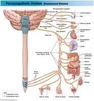
Types of Parasympathetic Ganglia
Terminal ganglia: Located near the target organ; usually paired.
Intramural ganglia: Embedded within the tissues of the target organ; consist of interconnected masses and clusters of ganglion cells.
Major Effects of Parasympathetic Activation
Constriction of pupils and focusing on near objects
Secretion by digestive glands
Secretion of hormones for nutrient absorption
Changes in blood flow and glandular activity during sexual arousal
Increased smooth muscle activity in the digestive tract
Stimulation of urination and defecation
Constriction of respiratory passageways
Reduction in heart rate and force of contraction
Parasympathetic Neurotransmitters and Receptors
Cholinergic Receptors
Nicotinic receptors: Located on postganglionic neurons of both sympathetic and parasympathetic divisions, and at neuromuscular junctions in the SNS. These are chemically gated Na+ channels; activation by ACh causes excitation of the postganglionic neuron.
Regulation of Autonomic Functions
Visceral Reflexes
Long reflexes: Coordinate the activities of entire organs. Visceral sensory neurons deliver information to the CNS, which processes it and sends motor commands to visceral effectors via the ANS. These reflexes are crucial for regulating internal organ activities.
Effects of Aging on the Nervous System
Anatomical and Physiological Changes
Changes begin by age 30 and accumulate over time.
85% of people over age 65 experience changes in mental performance and CNS function.
Reduction in brain size and weight, especially in the cerebral cortex (narrower gyri, wider sulci, larger subarachnoid space).
Reduction in the number of neurons (mainly cortical neurons; brainstem nuclei are spared).
Decreased blood flow to the brain due to arteriosclerosis, increasing the risk of stroke (CVA).
Loss of synaptic connections and decreased neurotransmitter production.
Functional Changes
Memory consolidation and retrieval become more difficult.
Sensory functions (hearing, balance, vision, smell, taste) decline.
Slower reaction times and weakened reflexes.
Decreased precision and speed of motor control.
Senile Dementia and Alzheimer’s Disease
Most elderly individuals retain functional independence, but some develop senile dementia (senility), characterized by memory loss, anterograde amnesia, and emotional disturbances.
Alzheimer’s disease is the most common form of senile dementia.
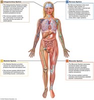
Integration of the Nervous System with Other Body Systems
The nervous system interacts with all other organ systems, regulating and integrating their functions to maintain homeostasis. For example, it controls muscle contraction, glandular secretion, and coordinates responses to internal and external stimuli.