 Back
The Autonomic Nervous System: Structure, Function, and Homeostatic Mechanisms
Study Guide - Smart Notes
 Tailored notes based on your materials, expanded with key definitions, examples, and context.
The Autonomic Nervous System (ANS)
Introduction to the ANS
The autonomic nervous system (ANS) is a division of the peripheral nervous system responsible for regulating involuntary physiological functions, including heart rate, blood pressure, digestion, and glandular activity. It operates largely below the level of consciousness to maintain homeostasis and adapt the body to changing internal and external environments.
Effectors: Innervates smooth muscle, cardiac muscle, and glands.
Function: Adjusts body functions such as blood flow, heart rate, and digestion to support body activities.
Control: Operates via subconscious (involuntary) control.
Alternate Names: Involuntary nervous system, general visceral motor system.
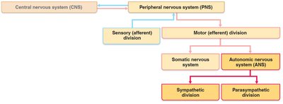
Somatic Nervous System vs. Autonomic Nervous System
Key Differences
The somatic and autonomic nervous systems both contain motor fibers but differ in their effectors, pathways, and neurotransmitter actions.
Effectors: Somatic NS targets skeletal muscles; ANS targets cardiac muscle, smooth muscle, and glands.
Efferent Pathways: Somatic NS uses a single neuron from CNS to muscle; ANS uses a two-neuron chain (preganglionic and postganglionic neurons).
Neurotransmitters: Somatic NS releases acetylcholine (ACh), always stimulatory. ANS preganglionic fibers release ACh; postganglionic fibers release ACh or norepinephrine (NE), which can be stimulatory or inhibitory depending on the receptor.
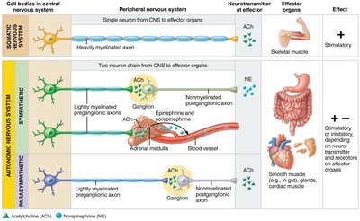
Overlap of Somatic and Autonomic Function
Integration and Coordination
Higher brain centers regulate and coordinate both somatic and autonomic systems. Many nerves contain both somatic and autonomic fibers, allowing for integrated responses to stimuli.
Adaptations often involve both skeletal muscles and visceral organs (e.g., increased heart rate during exercise).
Divisions of the ANS
Parasympathetic and Sympathetic Divisions
The ANS is divided into two main branches with generally opposing effects:
Parasympathetic Division: Promotes maintenance functions and conserves energy ("rest-and-digest").
Sympathetic Division: Mobilizes the body during activity ("fight-or-flight").
Dual Innervation: Most organs receive input from both divisions, which work antagonistically to maintain homeostasis.
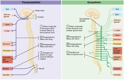
Parasympathetic Division
Structure and Function
The parasympathetic division is also called the craniosacral division because its fibers originate in the brainstem and sacral spinal cord. It is responsible for conserving energy and promoting housekeeping functions during rest.
Long preganglionic fibers extend from the CNS almost to target organs.
Major cranial nerves involved: Oculomotor (III), Facial (VII), Glossopharyngeal (IX), and Vagus (X).
Vagus nerve (X): Accounts for ~90% of all preganglionic parasympathetic fibers, serving thoracic and abdominal viscera.
Sacral outflow: Originates from S2–S4, serving pelvic organs and the distal half of the large intestine.
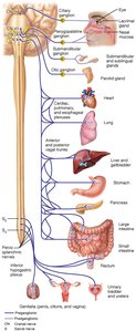
Sympathetic Division
Structure and Function
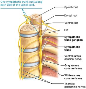
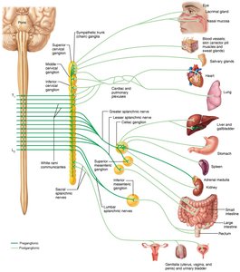
The sympathetic division is also called the thoracolumbar division, with fibers originating from the thoracic and lumbar regions of the spinal cord. It prepares the body for emergency or vigorous activity.
Short preganglionic and long postganglionic fibers.
Sympathetic trunk (chain): Contains 23 paravertebral ganglia (3 cervical, 11 thoracic, 4 lumbar, 4 sacral, 1 coccygeal).
Some fibers directly innervate the adrenal medulla, causing the release of epinephrine and norepinephrine into the blood.
Innervates more organs than the parasympathetic division, including structures only innervated by sympathetic fibers (e.g., sweat glands, arrector pili muscles, blood vessels).
Visceral Sensory Neurons and Reflexes
Visceral Sensory Neurons
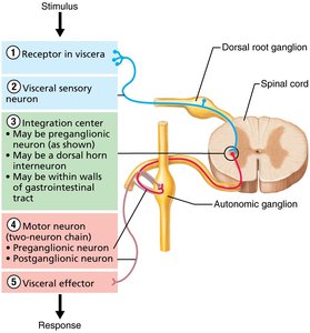
Visceral sensory neurons monitor internal conditions such as chemical changes, stretch, temperature, and irritation in the viscera. Their cell bodies are located in dorsal root ganglia and cranial nerve sensory ganglia.
Axons often travel alongside autonomic motor fibers.
Play a role in referred pain due to convergence on the same second-order sensory neurons as somatic sensory neurons.
Visceral Reflexes
Visceral reflex arcs are similar to somatic reflex arcs but have key differences:
Two consecutive neurons in the motor pathway.
Afferent fibers are visceral sensory neurons.
Effectors are smooth muscle, cardiac muscle, and glands.
Examples: Reflexes that empty the rectum and bladder.
Neurotransmitters and Receptors in the ANS
Major Neurotransmitters
The main neurotransmitters of the ANS are acetylcholine (ACh) and norepinephrine (NE).
Cholinergic fibers: Release ACh (all preganglionic axons, all parasympathetic postganglionic axons).
Adrenergic fibers: Release NE (most sympathetic postganglionic axons).
Cholinergic Receptors
Nicotinic receptors: Found on all postganglionic neurons, adrenal medulla cells, and skeletal muscle cells. Always stimulatory.
Muscarinic receptors: Found on all effector cells stimulated by postganglionic cholinergic fibers. Can be inhibitory or excitatory depending on the target organ.
Adrenergic Receptors
Alpha (α) receptors: Subclasses α1, α2.
Beta (β) receptors: Subclasses β1, β2, β3.
Effects depend on the receptor subtype and target organ (e.g., NE binding to β1 increases heart rate; epinephrine binding to β2 causes bronchial relaxation).
Parasympathetic and Sympathetic Interactions
Dual Innervation and Dynamic Antagonism
Most visceral organs receive input from both divisions, which interact antagonistically to maintain precise control over organ function.
Sympathetic division increases heart and respiratory rates, inhibits digestion and elimination.
Parasympathetic division decreases heart and respiratory rates, allows for digestion and waste elimination.
Some organs exhibit cooperative effects (e.g., sexual function).
Unique Roles of the Sympathetic Division
Regulates adrenal medulla, sweat glands, arrector pili muscles, kidneys, and most blood vessels (no parasympathetic innervation).
Thermoregulatory responses, renin release, and metabolic effects (increased metabolic rate, blood glucose, fat mobilization).
Localized vs. Diffuse Effects
Parasympathetic effects are short-lived and localized (ACh rapidly destroyed by acetylcholinesterase).
Sympathetic effects are longer-lasting and body-wide (NE inactivated slowly; adrenal medulla hormones prolong effects).
Effects of the Parasympathetic and Sympathetic Divisions on Various Organs
Organ System Effects
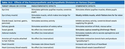
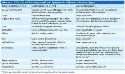
The following tables summarize the effects of parasympathetic and sympathetic stimulation on various organs:
Target Organ or System | Parasympathetic Effects | Sympathetic Effects |
|---|---|---|
Eye (iris) | Stimulates sphincter pupillae muscles; constricts pupils | Stimulates dilator pupillae muscles; dilates pupils |
Glands (nasal, lacrimal, gastric, pancreatic) | Stimulates secretory activity | Inhibits secretory activity; constricts blood vessels |
Salivary glands | Stimulates secretion of watery saliva | Stimulates copious sweating (cholinergic fibers) |
Adrenal medulla | No effect (no innervation) | Stimulates medulla cells to secrete epinephrine and norepinephrine |
Heart (muscle) | Decreases rate (slows heart) | Increases rate and force of heartbeat |
Lungs | Constricts bronchioles | Dilates bronchioles |
Digestive tract organs | Increases motility (peristalsis) and amount of secretion by digestive organs; relaxes sphincters to allow foodstuffs to move through tract | Decreases activity of glands and muscles of digestive system; constricts sphincters |
Liver | Increases glucose uptake from blood | Stimulates release of glucose to blood |
Kidney | No effect (no innervation) | Promotes renin release; causes vasoconstriction; decreases urine output |
Penis | Causes erection (vasodilation) | Causes ejaculation |
Blood vessels | Little or no effect (except vasodilation of vessels supplying external genitalia) | Constriction most vessels; increases blood pressure; diverts blood to muscles, brain, and heart |
Control of ANS Function
CNS Centers Involved
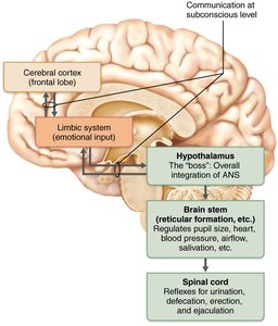
The ANS is regulated by several central nervous system centers:
Hypothalamus: Main integrative center for ANS activity; controls heart activity, blood pressure, body temperature, water balance, and endocrine activity.
Brain Stem: Reticular formation exerts direct influence; medullary centers regulate heart rate, blood vessel diameter, and gastrointestinal activities; midbrain controls pupil and lens muscles.
Spinal Cord: Controls defecation and micturition reflexes, subject to conscious override.
Cerebral Cortex: Can influence ANS activity via connections with the limbic system and hypothalamus; voluntary control possible through biofeedback.
Summary Table: Key Differences Between Parasympathetic and Sympathetic Divisions
Feature | Parasympathetic Division | Sympathetic Division |
|---|---|---|
Origin | Craniosacral (brainstem and sacral spinal cord) | Thoracolumbar (thoracic and lumbar spinal cord) |
Fiber Lengths | Long preganglionic, short postganglionic | Short preganglionic, long postganglionic |
Location of Ganglia | In or near effector organs | Close to spinal cord (sympathetic trunk) |
Neurotransmitters | ACh (preganglionic and postganglionic) | ACh (preganglionic), NE (postganglionic) |
General Function | Rest-and-digest, energy conservation | Fight-or-flight, energy mobilization |
