 Back
The Autonomic Nervous System: Structure, Function, and Integration
Study Guide - Smart Notes
 Tailored notes based on your materials, expanded with key definitions, examples, and context.
The Autonomic Nervous System (ANS)
Overview of the ANS
The autonomic nervous system (ANS) is a division of the peripheral nervous system responsible for regulating involuntary physiological processes, including heart rate, blood pressure, respiration, digestion, and sexual arousal. It maintains homeostasis by controlling smooth muscle, cardiac muscle, glands, and adipose tissue.
Homeostatic adjustment: Regulates water/electrolyte balance, nutrient/gas concentrations.
Coordination: Integrates cardiovascular, respiratory, digestive, urinary, and reproductive functions.
Effectors: Smooth muscle, cardiac muscle, glands, adipocytes.
Control center: Visceral motor nuclei in the hypothalamus.
Transmission: Two-neuron chain (preganglionic and postganglionic neurons) synapsing in autonomic ganglia.
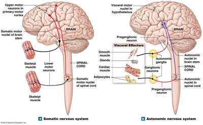
Comparison: Somatic vs. Autonomic Nervous System
The somatic nervous system (SNS) controls voluntary movements via a single lower motor neuron from the CNS to skeletal muscle. In contrast, the ANS uses a two-neuron chain to reach visceral effectors.
SNS: One neuron, effectors are skeletal muscles, control center in the primary motor cortex.
ANS: Two neurons, effectors are smooth/cardiac muscle, glands, adipocytes, control center in hypothalamus.
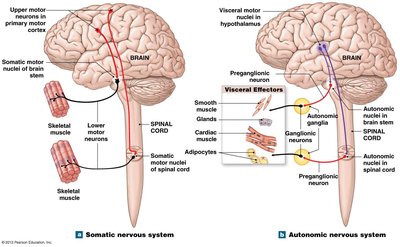
Organization of the ANS
Autonomic Ganglia and Neurons
Autonomic ganglia are clusters of neuronal cell bodies in the peripheral nervous system where preganglionic neurons synapse with postganglionic neurons. They are located adjacent to the spinal cord, near, or within effector organs.
Preganglionic neuron: Cell body in lateral gray horn or brainstem; axon extends to ganglion.
Postganglionic neuron: Cell body in autonomic ganglion; axon extends to effector.
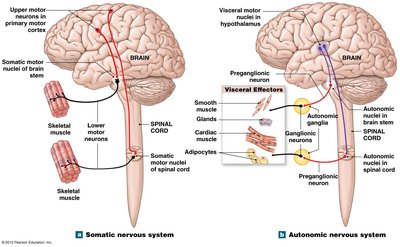
Divisions of the ANS
Sympathetic Division
The sympathetic division prepares the body for heightened somatic activity ("fight or flight").
Origin: Thoracic and lumbar spinal nerves (T1-L2; thoracolumbar).
Ganglia: Near the spinal cord (sympathetic chain, collateral ganglia, adrenal medullae).
Generalized response: Increased alertness, energy, and metabolic rate.
Parasympathetic Division
The parasympathetic division supports "rest and repose" functions, promoting visceral activity and energy conservation.
Origin: Cranial nerves III, VII, IX, X and sacral spinal nerves (S2-S4; craniosacral).
Ganglia: Near or within effector organs (terminal and intramural ganglia).
Generalized response: Promotes digestion, nutrient absorption, and energy storage.
Enteric Nervous System (ENS)
The enteric nervous system is a network of neurons in the digestive tract wall, capable of local reflexes and modulated by the ANS.
Structures of the Sympathetic Division
Preganglionic Neurons and Pathways
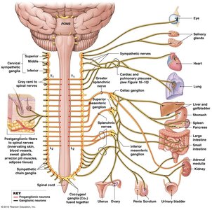
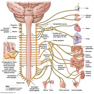
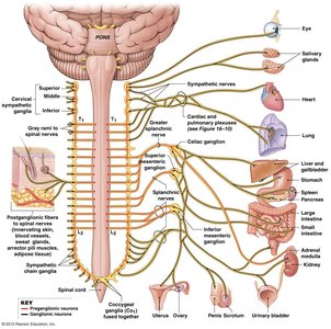
Sympathetic preganglionic neurons originate in the lateral gray horns of T1-L2 and project through ventral roots, white rami communicantes, and various nerves to reach ganglia.
Sympathetic chain ganglia: Paired, lateral to vertebral column; innervate body wall, head, limbs, thoracic cavity.
Collateral ganglia: Unpaired, anterior to vertebral bodies; innervate abdominopelvic organs.
Adrenal medullae: Paired glands above kidneys; neuroendocrine cells secrete epinephrine and norepinephrine.
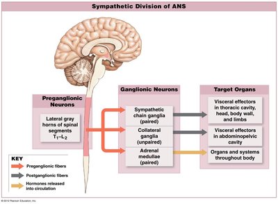
Sympathetic Pathways
There are four main sympathetic pathways:
Spinal nerves: To body wall and limbs (synapse in chain ganglia, postganglionic fibers via gray rami communicantes).
Sympathetic nerves: To head and thoracic cavity (postganglionic fibers extend through sympathetic nerves).
Splanchnic nerves: To abdominopelvic cavity (preganglionic fibers synapse in collateral ganglia).
Bloodstream: Via adrenal medullae (neuroendocrine cells release hormones into blood).
Sympathetic Activation
Sympathetic activation is a coordinated response during physiological crisis, controlled by the hypothalamus and involving hormone release from the adrenal medullae.
Increased alertness, energy, and euphoria
Elevated blood pressure, heart rate, and ventilation
Mobilization of energy reserves
Structures of the Parasympathetic Division
Preganglionic Neurons and Ganglia
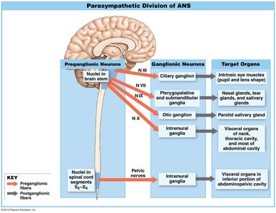
Parasympathetic preganglionic neurons originate in cranial nerve nuclei (III, VII, IX, X) and sacral spinal cord (S2-S4). Their fibers synapse in terminal or intramural ganglia near or within target organs.
Terminal ganglia: Adjacent to target organ
Intramural ganglia: Embedded within target organ tissues
Ganglia Associated with Cranial Nerves
Oculomotor (III): Ciliary ganglion (eye)
Facial (VII): Pterygopalatine (tears), submandibular (saliva)
Glossopharyngeal (IX): Otic ganglion (saliva)
Vagus (X): Intramural ganglia in visceral organs
Parasympathetic Activation
Parasympathetic activation supports rest, digestion, and energy conservation.
Pupil constriction, near vision focusing
Promotion of digestive secretion and motility
Nutrient absorption and utilization
Urinary bladder contraction
Constriction of respiratory passages
Reduction in heart rate and contractile force
Sexual arousal
Neurotransmission in the ANS
Synapses and Neurotransmitters
ANS synapses are classified by the neurotransmitter released:
Cholinergic: Release acetylcholine (ACh); effects are excitatory and brief.
Adrenergic: Release norepinephrine (NE); effects are longer-lasting.
Nitroxidergic: Release nitric oxide (NO); relaxes smooth muscle in blood vessels.
In the sympathetic division, preganglionic to ganglionic synapses are cholinergic; ganglionic to effector synapses are usually adrenergic, but may also be cholinergic or nitroxidergic. In the parasympathetic division, all synapses are cholinergic.
Receptors of the ANS
Cholinergic receptors: Detect ACh; include nicotinic (on ganglionic neurons, chemically gated channels) and muscarinic (on effector cells, G protein-coupled).
Adrenergic receptors: Detect E/NE; include alpha (α) and beta (β) classes with various subtypes and effects.
Integration and Control of ANS Activity
Coordination of ANS Divisions
Most organs receive dual innervation from both sympathetic and parasympathetic divisions, which usually exert opposite effects. Sympathetic effects are more pervasive due to adrenal hormone release.
Autonomic plexuses: Nerve networks in thoracic and abdominopelvic cavities containing both sympathetic and parasympathetic fibers (e.g., cardiac, pulmonary, celiac plexuses).
Autonomic tone: Baseline level of spontaneous activity in ANS neurons, allowing bidirectional adjustment.
Visceral Reflexes
Visceral reflexes are automatic neural responses involving ANS motor neurons. All are polysynaptic and can be modified by the hypothalamus.
Long reflex: Involves CNS interneurons; controls entire organs.
Short reflex: Involves ganglionic interneurons; controls small regions within organs.
Higher Levels of Autonomic Control
Reflex centers in the medulla oblongata control individual autonomic reflexes, influenced by hypothalamic centers, which integrate input from the limbic system, thalamus, and cerebral cortex.
Integration of SNS and ANS Function
The somatic and autonomic nervous systems can be coordinated by reflexes or higher brain commands. The limbic system links the cerebral cortex (SNS control) and hypothalamus (ANS control), allowing conscious influence over autonomic responses (e.g., calming oneself before an exam).
