 Back
BackChapter 17- The Autonomic Nervous System: Structure, Function, and Divisions
Study Guide - Smart Notes
 Tailored notes based on your materials, expanded with key definitions, examples, and context.
Autonomic Nervous System (ANS)
Overview of the ANS
The Autonomic Nervous System (ANS) is a division of the peripheral nervous system that regulates involuntary physiological processes, including heart rate, blood pressure, respiration, digestion, and glandular activity. It innervates adipose tissue, smooth muscle, cardiac muscle, and glands. The ANS operates via a two-neuron efferent pathway: a preganglionic fiber and a postganglionic fiber.
Preganglionic fiber: Originates in the CNS and synapses in a ganglion.
Postganglionic fiber: Extends from the ganglion to the effector organ.
In contrast, the Somatic Nervous System controls voluntary movements of skeletal muscle and uses a single motor neuron in its efferent pathway.
Divisions of the ANS
Sympathetic and Parasympathetic Divisions
The ANS is divided into two main subdivisions, each with distinct anatomical and functional characteristics:
Sympathetic Division: Prepares the body for 'fight-or-flight' responses during stress or activity.
Parasympathetic Division: Promotes 'rest-and-digest' activities during periods of rest and recovery.
Enteric Nervous System
The Enteric Nervous System is a complex network of neurons within the walls of the digestive tract. It can function independently of the CNS to coordinate local reflexes related to digestion.
Sympathetic Division
Anatomy and Pathways
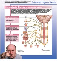
The sympathetic division has three main locations for its ganglia:
Sympathetic chain ganglia: Located on both sides of the vertebral column.
Collateral ganglia: Positioned anterior to the vertebral column; postganglionic fibers from these ganglia innervate abdominopelvic organs.
Adrenal medulla: The inner region of the adrenal gland, which releases hormones into the bloodstream.
Preganglionic neurons originate in the thoracic and lumbar regions of the spinal cord, a pattern known as thoracolumbar outflow.
Neurotransmitters and Receptors
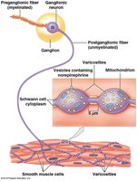
Preganglionic fibers: Release acetylcholine (ACh), a cholinergic neurotransmitter, which excites ganglionic neurons.
Postganglionic fibers: Release norepinephrine (NE), an adrenergic neurotransmitter, which can have excitatory or inhibitory effects depending on the receptor type (alpha or beta) on the effector cell.
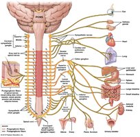
Anatomical Distribution
Sympathetic postganglionic fibers are distributed to various organs, including the heart, lungs, digestive organs, and blood vessels. Splanchnic nerves serve abdominopelvic organs.
Adrenal Medulla
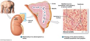
The adrenal medulla is a modified sympathetic ganglion. When stimulated by sympathetic preganglionic fibers, it secretes epinephrine and norepinephrine into the bloodstream, acting as hormones to produce widespread effects.
Effects of Sympathetic Stimulation
Increased alertness and energy
Elevated heart rate, blood pressure, and respiratory rate
Increased muscle tone
Mobilization of energy reserves (glucose and lipids)
Sympathetic Motor Pathways
Sympathetic motor pathways involve the release of ACh at the ganglion and NE at the effector. The effect on the target cell depends on the receptor type (alpha or beta).
Parasympathetic Division
Anatomy and Pathways
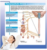
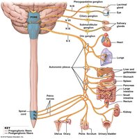
Parasympathetic ganglia are located within or near the target organs, resulting in more specific and localized effects. Preganglionic neurons originate in the brainstem and sacral spinal cord, known as craniosacral outflow.
Neurotransmitters and Receptors
Preganglionic fibers: Release ACh (cholinergic), exciting ganglionic neurons.
Postganglionic fibers: Also release ACh, which can excite or inhibit the effector depending on the receptor type:
Nicotinic receptors: Always excitatory; found on ganglionic neurons and skeletal muscle.
Muscarinic receptors: Can be excitatory or inhibitory; found on parasympathetic target organs.
Effects of Parasympathetic Stimulation
Pupil constriction
Stimulation of digestive gland secretion
Promotion of nutrient absorption
Increased smooth muscle activity in the digestive tract
Defecation and urination
Constriction of airways
Reduced heart rate and contraction force
Sexual arousal and stimulation
Anatomical Distribution
Parasympathetic output is distributed to organs such as the eye, salivary glands, heart, lungs, digestive organs, and reproductive organs.
Dual Innervation
Antagonistic Effects
Most vital organs receive input from both sympathetic and parasympathetic divisions, often producing opposite effects. For example, the sympathetic division increases heart rate, while the parasympathetic division decreases it.
Comparison of Sympathetic and Parasympathetic Divisions
Key Differences
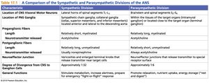
The following table summarizes the main differences between the sympathetic and parasympathetic divisions of the ANS:
Characteristic | Sympathetic Division | Parasympathetic Division |
|---|---|---|
Location of CNS Visceral Motor Neurons | Lateral horns of spinal segments T1–L2 | Brainstem and spinal segments S2–S4 |
Location of PNS Ganglia | Sympathetic chain, collateral ganglia, adrenal medulla | Near or within target organs |
Preganglionic Fiber Length | Relatively short, myelinated | Relatively long, myelinated |
Postganglionic Fiber Length | Relatively long, unmyelinated | Relatively short, unmyelinated |
Neurotransmitter Released (Preganglionic) | Acetylcholine (ACh) | Acetylcholine (ACh) |
Neurotransmitter Released (Postganglionic) | Norepinephrine (NE), sometimes ACh or NO | Acetylcholine (ACh) |
General Functions | Stimulates metabolism, increases alertness, prepares for emergency ('fight or flight') | Promotes relaxation, nutrient uptake, energy storage ('rest and digest') |
Visceral Reflexes
Autonomic Reflexes
Visceral reflexes are automatic, unconscious responses to stimulation of visceral organs. Many are coordinated locally within the organ system, sometimes bypassing the central nervous system entirely.
