 Back
BackThe Autonomic Nervous System: Structure, Function, and Neurotransmission
Study Guide - Smart Notes
 Tailored notes based on your materials, expanded with key definitions, examples, and context.
Autonomic Nervous System (ANS)
Overview of the Autonomic Nervous System
The Autonomic Nervous System (ANS) is a division of the peripheral nervous system that regulates involuntary physiological processes, including heart rate, blood pressure, respiration, digestion, and sexual arousal. It operates largely below the level of consciousness and is essential for maintaining homeostasis.
Somatic Nervous System: Controls voluntary movements of skeletal muscles.
Autonomic Nervous System: Controls involuntary actions of smooth muscle, cardiac muscle, and glands.
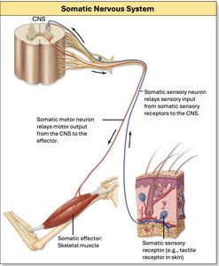
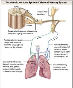
General Anatomic Organization of the ANS
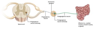
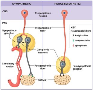
The ANS consists of a two-neuron chain: a preganglionic neuron (cell body in the CNS) and a postganglionic neuron (cell body in an autonomic ganglion). The postganglionic neuron innervates the target organ.
Preganglionic neuron: Cell body in the CNS; axon projects to an autonomic ganglion. Type B neuron = myelinated
Postganglionic neuron: Cell body in the autonomic ganglion; axon projects to the effector (smooth muscle, cardiac muscle, or gland). Type C neuron = unmyelinated
Divisions of the Autonomic Nervous System
Sympathetic Division
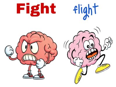
The sympathetic division is responsible for the "fight or flight" response, preparing the body for stressful or emergency situations. It increases alertness, metabolic rate, and energy availability.
Active during high stress or emergency situations.
Allows for maximum energy expenditure.
Originates from the thoracolumbar region (T1–L2) of the spinal cord.
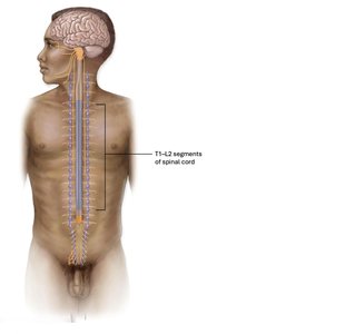
Layout of the Sympathetic Division
Short, branching preganglionic neurons = Type B
Long postganglionic neurons = Type C
Autonomic ganglia are close to the spinal cord
Types of Sympathetic Ganglia
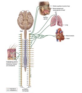
Sympathetic Chain Ganglia: Lateral to the spinal cord; innervate head, body wall, limbs, and thoracic cavity.
Eyes = contracts dialator pupillae muscle, relaxes ciliary muscles for distant vision
Heart = how your heart feels when your are stressed, increased heart rate, force of contraction
Lungs = bronchodilation, increase in rate of respiration
Other effects:
increased metabolic rate and energy use
increased mental alterness
increased sweating
decreased blood flow to the skin
increased blood flow to skeletal muscles
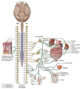
Collateral Ganglia: Anterior to the vertebral column; innervate abdominopelvic organs.
Celiac ganglion = stomach, gallbladder, liver, pancreas, and spleen
Superior mesenteric ganglion = small intestine and proximal 2/3 large interstine
Inferior mesenteric ganglion = distal 1/3 of large intestine, kidney, bladder, and reproductive organs
Digestive system
decreased digestive secretions
increased glycogen breakdown
contract sphincters in GI tract
relax smooth muscle in GI tract
Urinary system
decreased blood flow to urinary organs
contract urinary sphincters
relax smooth muscle
Reproductive system
ejaculation in males
uterine contraction in females
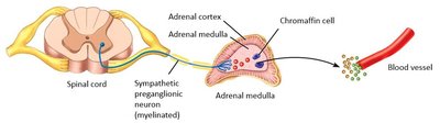
Adrenal Medulla: Center of the adrenal gland; releases hormones into the blood for a widespread effect.
Chromaffin cells = take the place of the postganglionic neuron and release epinephrine and norepinesphrine (as hormones)
neurotransmitters released to the blood -> provide sympathetic innervation to the entire body all at once, has more generalized effect
Parasympathetic Division
The parasympathetic division is responsible for the "rest and digest" response, promoting maintenance activities and conserving body energy. It is dominant during resting conditions.
Promotes digestion, energy storage, and tissue repair.
Originates from the craniosacral region (brainstem and S2–S4 spinal segments).

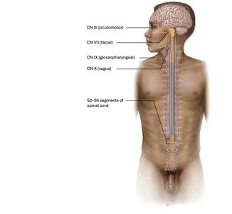
Layout of the Parasympathetic Division
Long, unbranched preganglionic neurons
Short postganglionic neurons
Autonomic ganglia are close to or within the target tissue
Types of Parasympathetic Ganglia
Terminal Ganglia: Located near the target organ
Intramural Ganglia: Located within the wall of the target organ
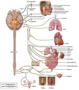
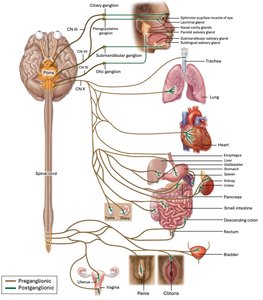
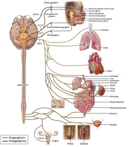
Ciliary Ganlgion
oculomoter nerve is preganglionic
innervates the smooth muscle in the eyes
sphrincter pupillae
contract ciliary muscles = visual accommodation
Pterygopalatine Ganglion
facial nerve is preganglionic
innervates nasal and lacrimal glands
increased secretion in secretion in both types. ofgland
Submandibular Ganglion
facial nerve is preganglionic
innervates the submanidbular and sublingual salivary gland
increases salivary secretion
Optic Ganglion
glossopharyngeal nerve is preganglionic
innervates parotid salivary glands
increases salivary secretion
intramural Ganglia
vagus nerve is preganglioninc
cardiac muscle cells = decreases heart rate and force of contraction
smooth muscle in airways = bronchoconstriction and decreased rate of respiration
digestive organs
smooth muscle in GI tract = increases motility
increases secretion from digestive glands
relaxes sphincters in GI tract
Intramural Ganglia
pelvic nerves are preganlgionic
urinary tract
increases urine production
relaxes urinary sphincters
rectum = relaxes sphincters
reproductive organs
increases gametogenesis = makes more sperm/eggs
causes erections in males
other parasympathetic effects
decreased sweating
decreased mental altertness
Enteric Division
The enteric division is a network of neurons within the walls of the digestive tract that regulates gastrointestinal function independently but is modulated by the sympathetic and parasympathetic divisions.
Provides localized control of digestive activity
Coordinates reflexes for peristalsis and secretion
Neurotransmitters and Receptors in the ANS
Sympathetic Neurotransmitters and Receptors
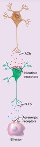
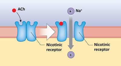
Preganglionic neurons release acetylcholine (ACh), which binds to nicotinic receptors on postganglionic neurons.
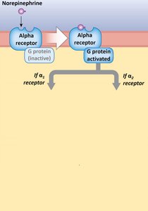
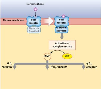
Postganglionic neurons release norepinephrine (N.Epi) or epinephrine (Epi), which bind to adrenergic receptors (alpha and beta types) on target cells.
Receptor Type | Main Effect | Location |
|---|---|---|
α1 | Excitatory (smooth muscle contraction, gland secretion) | Blood vessels, glands |
α2 | Inhibitory (decreased cAMP, closes GI sphincters) | GI tract sphincters |
β1 | Stimulates cardiac muscle, increases metabolism | Heart |
β2 | Relaxes smooth muscle (bronchodilation, vasodilation) | Lungs, blood vessels |
β3 | Increases lipolysis | Adipose tissue |
Parasympathetic Neurotransmitters and Receptors
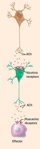
Preganglionic neurons release acetylcholine (ACh), which binds to nicotinic receptors on postganglionic neurons.
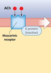
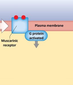
Postganglionic neurons release acetylcholine (ACh), which binds to muscarinic receptors on target cells.
Receptor Type | Main Effect | Location |
|---|---|---|
Nicotinic | Excitatory (depolarization) | All postganglionic neurons |
Muscarinic | Excitatory or inhibitory (depending on target) | Effector organs (heart, smooth muscle, glands) |
Medications and Adrenergic Receptors
many medications act by binding to adrenergic receptors
Blockers = block the receptor site and prevent N.epi binding
Agonists = Mimic effects of N. Epi binding
Dual Innervation and Autonomic Tone
Dual Innervation
Most organs receive input from both sympathetic and parasympathetic divisions, which typically have opposing effects. This allows for precise regulation of organ function.
Autonomic Tone
Both divisions of the ANS are partially active at all times, creating a baseline level of activity known as autonomic tone. The balance between sympathetic and parasympathetic tone determines the overall effect on an organ.
Sympathetic tone: Maintains partial constriction of blood vessels, helping to regulate blood pressure.
Parasympathetic tone: Maintains resting heart rate at about 72 bpm.
Summary Table: Comparison of Somatic and Autonomic Nervous Systems
Feature | Somatic Nervous System | Autonomic Nervous System |
|---|---|---|
Control | Voluntary | Involuntary |
Effector Organs | Skeletal muscle | Cardiac muscle, smooth muscle, glands |
Number of Neurons | One (CNS to effector) | Two (preganglionic and postganglionic) |
Neurotransmitter | Acetylcholine (ACh) | ACh, norepinephrine (N.Epi), epinephrine (Epi) |
Effect | Always excitatory | Excitatory or inhibitory |
