 Back
Ch. 14 The Autonomic Nervous System: Structure, Function, and Clinical Relevance
Study Guide - Smart Notes
 Tailored notes based on your materials, expanded with key definitions, examples, and context.
The Autonomic Nervous System (ANS)
Introduction to the ANS
The autonomic nervous system (ANS) is a division of the peripheral nervous system responsible for regulating involuntary physiological processes, including heart rate, blood pressure, respiration, digestion, and glandular activity. It operates largely below the level of consciousness and is essential for maintaining homeostasis.
Effectors: Smooth muscle, cardiac muscle, and glands
Control: Subconscious (involuntary)
Other names: Involuntary nervous system, visceral motor system
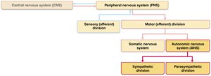
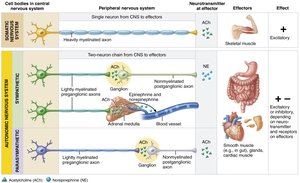
Comparison: Somatic vs. Autonomic Nervous Systems
Key Differences
Although both systems have motor fibers, they differ in their effectors, neural pathways, and neurotransmitter actions.
Somatic Nervous System (SNS): Innervates skeletal muscles; single, thick, myelinated axon from CNS to muscle; always excitatory (acetylcholine, ACh).
Autonomic Nervous System (ANS): Innervates cardiac muscle, smooth muscle, and glands; two-neuron chain (preganglionic and postganglionic); can be excitatory or inhibitory (ACh or norepinephrine, NE).
Divisions of the ANS
Parasympathetic vs. Sympathetic Divisions
The ANS is divided into two main branches that typically have opposing effects on target organs, maintaining homeostasis through dynamic antagonism.
Parasympathetic Division: "Rest-and-digest"; conserves energy, promotes maintenance functions (e.g., digestion, urination, defecation).
Sympathetic Division: "Fight-or-flight"; mobilizes body during activity, increases heart rate, dilates pupils, inhibits digestion.
Dual Innervation: Most organs receive input from both divisions, which usually have opposite effects.
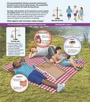
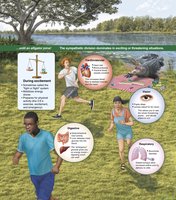
Key Anatomical Differences
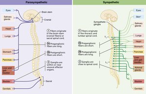
Sites of Origin: Parasympathetic fibers are craniosacral; sympathetic fibers are thoracolumbar.
Fiber Lengths: Parasympathetic has long preganglionic and short postganglionic fibers; sympathetic has short preganglionic and long postganglionic fibers.
Ganglia Location: Parasympathetic ganglia are near or within target organs; sympathetic ganglia are close to the spinal cord.
Parasympathetic Division
Organization and Pathways
The parasympathetic division is also called the craniosacral division. Its preganglionic neurons originate in the brainstem and sacral spinal cord, and synapse in terminal ganglia near or within target organs.
Cranial Outflow: Oculomotor, facial, glossopharyngeal (IX, activates parotid salivary glands), and vagus nerves (As the vagus nerves (X) pass into the thorax, they send branches to the cardiac plexuses supplying fibers to the heart that slow heart rate.)
Sacral Outflow: Pelvic splanchnic nerves to pelvic organs and distal large intestine
Sympathetic Division
Organization and Pathways
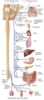
The sympathetic division is also called the thoracolumbar division. Preganglionic neurons originate in the thoracic and lumbar spinal cord, and synapse in sympathetic trunk (paravertebral) or collateral (prevertebral) ganglia; sympathetic nerve impulses act only briefly, they provoke hormonal effects that last much longer.
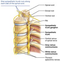
Sympathetic Trunks: Chains of ganglia flanking the vertebral column
Collateral Ganglia: Located anterior to the vertebral column (e.g., celiac, superior mesenteric)
Unique Targets: Sweat glands, arrector pili muscles, blood vessels (sympathetic only)
Sympathetic Pathways
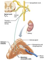
Preganglionic fibers may synapse at the same level, ascend/descend to another level, or pass through to collateral ganglia (cannot synapse in the sacral region). Some fibers pass directly to the adrenal medulla, stimulating hormone release.
Three Main Pathways: To body wall, head/thorax, and abdominal/pelvic organs
Adrenal Medulla: Chromaffin cells release epinephrine and norepinephrine into the blood
Visceral Reflexes
Visceral Reflex Arcs
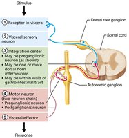
Visceral reflexes control involuntary functions such as heart rate and digestion. They involve sensory input from visceral organs, integration in the CNS, and motor output via the ANS.
Components: Receptor, sensory neuron, integration center, motor neuron, effector
Differences from Somatic Reflexes: Two-neuron motor pathway, visceral effectors
Neurotransmitters and Receptors
Major Neurotransmitters
Acetylcholine (ACh): Released by all preganglionic neurons and parasympathetic postganglionic neurons
Norepinephrine (NE): Released by most sympathetic postganglionic neurons
Receptor Types
Cholinergic Receptors: Bind ACh; include nicotinic (always excitatory) and muscarinic (excitatory or inhibitory depending on target)
Adrenergic Receptors: Bind NE or epinephrine; include alpha and beta subclasses with varying effects
Parasympathetic and Sympathetic Interactions
Dynamic Antagonism and Tone
Most organs receive dual innervation, allowing precise regulation. Sympathetic tone maintains blood pressure; parasympathetic tone dominates heart and digestive organs under resting conditions.
Antagonistic Effects: Sympathetic increases heart rate, inhibits digestion; parasympathetic decreases heart rate, promotes digestion
Cooperative Effects: Both divisions cooperate in reproductive functions (e.g., erection and ejaculation)
Central Control of the ANS
Hierarchy of Control
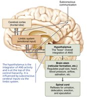
The ANS is regulated by CNS centers, primarily the hypothalamus (integrative center at the top of the autonomic-nervous-system control hierarchy), with input from the brainstem, spinal cord, and cerebral cortex (via the limbic system).
Hypothalamus: Main integrative center; directs both sympathetic and parasympathetic activity
Brainstem: Regulates heart rate, blood vessel diameter, gastrointestinal activities
Spinal Cord: Controls defecation, urination, and sexual reflexes
Disorders of the ANS
Common Disorders
Hypertension: Overactive sympathetic vasoconstriction; treated with adrenergic blockers
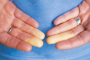
Raynaud’s Disease: Exaggerated vasoconstriction in fingers/toes; treated with vasodilators
Autonomic Dysreflexia: Uncontrolled activation of autonomic neurons, especially in spinal cord injury patients
Autonomic neuropathy is a common complication of diabetes mellitus.
Blood “shunting” helps maintain circulation to vital organs during circulatory shock.
Developmental Aspects of the ANS
Development and Aging
Origin: Preganglionic neurons from neural tube; postganglionic neurons and ganglia from neural crest
Aging: ANS efficiency declines with age, leading to issues such as constipation, dry eyes, and orthostatic hypotension
