 Back
Chapter 16- Lecture 1
Study Guide - Smart Notes
 Tailored notes based on your materials, expanded with key definitions, examples, and context.
The Autonomic Nervous System (ANS)
Overview of the Autonomic Nervous System
The autonomic nervous system (ANS) is a division of the peripheral nervous system that regulates involuntary physiological processes, including heart rate, blood pressure, respiration, digestion, and sexual arousal. It operates largely below the level of consciousness and is essential for maintaining homeostasis.
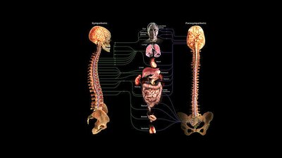
Divisions: The ANS is divided into the sympathetic and parasympathetic divisions, each with distinct anatomical and functional characteristics.
Comparison with Somatic Nervous System: The somatic nervous system controls voluntary movements of skeletal muscles, while the ANS controls involuntary actions of smooth muscle, cardiac muscle, and glands.
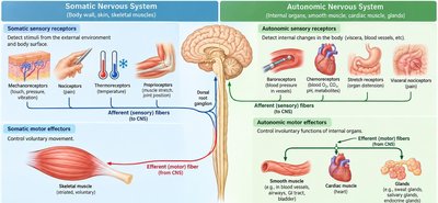
Comparison of Somatic and Autonomic Nervous Systems
The somatic and autonomic nervous systems differ in their effectors, neural pathways, neurotransmitters, and control mechanisms.
Somatic Nervous System: Controls voluntary movements via a single neuron pathway from the CNS to skeletal muscle. Neurotransmitter: acetylcholine (ACh).
Autonomic Nervous System: Controls involuntary functions via a two-neuron chain (preganglionic and postganglionic neurons). Neurotransmitters: acetylcholine (ACh) and norepinephrine (NE).
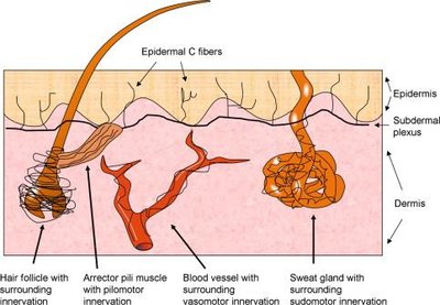
Visceral Effectors in the Skin
Visceral effectors in the skin include blood vessels, sweat glands, and arrector pili muscles. These structures are innervated by the autonomic nervous system and are responsible for thermoregulation, piloerection, and sweat production.
Blood vessels: Vasoconstriction and vasodilation regulate blood flow and temperature.
Sweat glands: Control sweating for thermoregulation.
Arrector pili muscles: Cause hair to stand up (goosebumps) in response to cold or emotional stimuli.
Organization of the Autonomic Nervous System
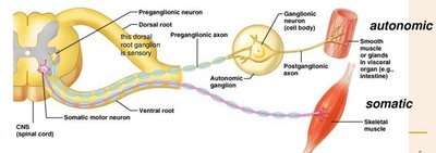
Neural Pathways: Somatic vs. Autonomic
The neural pathways of the somatic and autonomic nervous systems differ in the number of neurons and the location of synapses.
Somatic Pathway: Single motor neuron extends from the CNS to the effector muscle.
Autonomic Pathway: Involves a preganglionic neuron (from CNS to autonomic ganglion) and a postganglionic neuron (from ganglion to effector organ).
Sites of Ganglia in the Autonomic Nervous System
Autonomic ganglia are sites where preganglionic neurons synapse with postganglionic neurons. There are three main locations:
Paravertebral (Sympathetic Chain) Ganglia: Located alongside the vertebral column.
Prevertebral (Collateral) Ganglia: Located anterior to the vertebral column, near major abdominal arteries.
Adrenal Medulla: Modified sympathetic ganglion where preganglionic fibers synapse directly on chromaffin cells, which release hormones into the bloodstream.
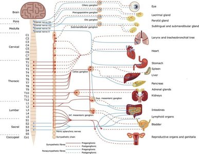
Divisions of the Autonomic Nervous System
Sympathetic (Thoracolumbar) Division
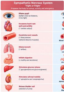
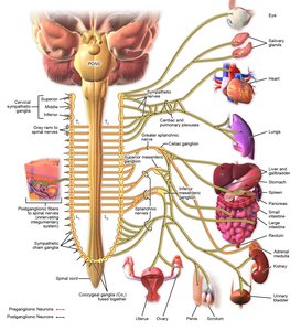
The sympathetic division prepares the body for 'fight or flight' responses. Its preganglionic neurons originate from the thoracic and lumbar regions (T1–L2) of the spinal cord.
Functions: Increases heart rate, dilates pupils, inhibits digestion, stimulates glucose release, and increases blood flow to muscles.
Pathways: Preganglionic fibers may synapse in sympathetic chain ganglia, collateral ganglia, or the adrenal medulla.
Parasympathetic (Craniosacral) Division
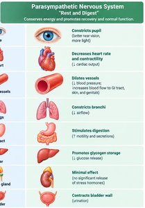
The parasympathetic division promotes 'rest and digest' activities. Its preganglionic neurons originate from the brainstem and sacral spinal cord (craniosacral outflow).
Functions: Decreases heart rate, constricts pupils, stimulates digestion, and promotes energy storage.
Pathways: Long preganglionic fibers synapse in ganglia close to or within target organs.
Functional Comparison: Sympathetic vs. Parasympathetic
Major Effects on Organs
The sympathetic and parasympathetic divisions often have opposing effects on the same organs, maintaining homeostasis through dynamic balance.
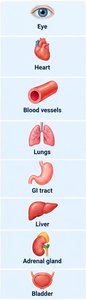
Organ | Sympathetic Effect | Parasympathetic Effect |
|---|---|---|
Pupil (Eye) | Dilates pupil | Constricts pupil |
Heart | Increases rate and force | Decreases rate |
Blood Vessels | Constriction (most) | Dilation (some) |
Lungs | Dilates bronchi | Constricts bronchi |
GI Tract | Inhibits motility/secretion | Stimulates motility/secretion |
Liver | Stimulates glucose release | Promotes glycogen storage |
Adrenal Gland | Stimulates hormone release | Minimal effect |
Bladder | Relaxes wall | Contracts wall |
Effects on Skin Structures
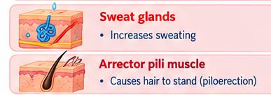
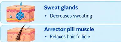
The ANS regulates sweat glands and arrector pili muscles in the skin, with distinct effects from each division.
Sympathetic: Increases sweating, causes piloerection (hair stands up).
Parasympathetic: Decreases sweating, relaxes hair follicles.
Neurotransmitters and Receptors
Somatic vs. Autonomic Neurotransmission
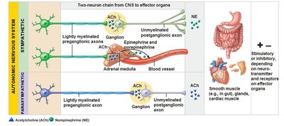
The somatic nervous system uses acetylcholine (ACh) at the neuromuscular junction, while the ANS uses both ACh and norepinephrine (NE) depending on the division and target organ.
Somatic: ACh stimulates skeletal muscle contraction.
Sympathetic: Preganglionic neurons release ACh; postganglionic neurons typically release NE (except sweat glands, which use ACh).
Parasympathetic: Both pre- and postganglionic neurons release ACh.

Sympathetic Pathways and Ganglia
Sympathetic Chain and Collateral Ganglia
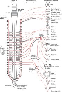
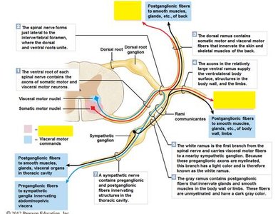
Sympathetic preganglionic fibers may synapse in the sympathetic chain ganglia (paravertebral), collateral ganglia (prevertebral), or directly on the adrenal medulla. These pathways allow for widespread and coordinated responses during stress.
Sympathetic Chain Ganglia: Innervate organs in the thoracic cavity, head, body wall, and limbs.
Collateral Ganglia: Innervate organs in the abdominopelvic cavity.
Adrenal Medulla: Secretes epinephrine and norepinephrine into the bloodstream for systemic effects.

Adrenal Medulla as a Modified Ganglion
The adrenal medulla acts as a modified sympathetic ganglion. Preganglionic sympathetic fibers synapse directly on chromaffin cells, which release epinephrine and norepinephrine into the bloodstream, producing prolonged and widespread sympathetic effects.
Hormones released: Epinephrine (adrenaline) and norepinephrine (noradrenaline).
Effect: Amplifies and sustains the fight-or-flight response.
Clinical and Functional Considerations
Effects of Sympathetic Trunk Damage
Damage to different regions of the sympathetic trunk can affect various organ systems:
Cervical/Thoracic Damage: May impair vision, salivation, thermoregulation, and stress responses.
Lumbar Damage: May affect bladder control, genital function, and blood flow to lower limbs.
Sample Clinical Questions
Pupil Dilation During Stress: Sympathetic fibers from T1–T2 via the superior cervical ganglion innervate the dilator pupillae muscle.
Sympathetic Trunk Lesions: May cause warmer, redder skin due to reduced vasoconstrictor activity.
Adrenal Medulla Innervation: Preganglionic sympathetic fibers synapse directly on adrenal medulla cells (not the cortex).
Summary Table: Somatic vs. Autonomic Nervous System
Feature | Somatic | Autonomic |
|---|---|---|
Effectors | Skeletal muscle | Cardiac muscle, smooth muscle, glands |
Control | Voluntary | Involuntary |
Neural Pathway | Single neuron from CNS | Two-neuron chain (pre- and postganglionic) |
Neurotransmitter | Acetylcholine (ACh) | ACh, norepinephrine (NE) |
Effect | Always excitatory | Excitatory or inhibitory |
