 Back
Chapter 16- Lecture 2
Study Guide - Smart Notes
 Tailored notes based on your materials, expanded with key definitions, examples, and context.
The Autonomic Nervous System (ANS)
Overview of the ANS
The autonomic nervous system (ANS) regulates involuntary physiological processes, including heart rate, blood pressure, respiration, digestion, and glandular activity. It is divided into the sympathetic and parasympathetic divisions, each with distinct anatomical and functional characteristics.
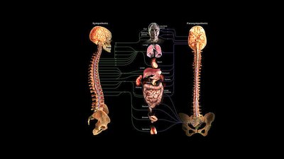
Sympathetic Division: Prepares the body for 'fight or flight' responses.
Parasympathetic Division: Promotes 'rest and digest' activities.
Sympathetic Neurotransmitters and Receptors
Neurotransmitter Release in the Sympathetic Division
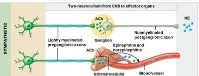
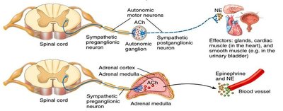
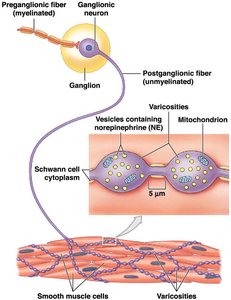
Sympathetic pathways typically involve a two-neuron chain from the central nervous system (CNS) to effector organs. The neurotransmitters released at each synapse determine the physiological response.
Preganglionic Neurons: Release acetylcholine (ACh) at synapses in autonomic ganglia or the adrenal medulla. This effect is always excitatory.
Postganglionic Neurons: Most release norepinephrine (NE) at target organs, but some (e.g., those innervating sweat glands and certain blood vessels) release ACh.
Special Cases: Cholinergic Sympathetic Neurons
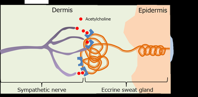
Sweat Glands: Sympathetic nerves release ACh, causing sweating and aiding thermoregulation.
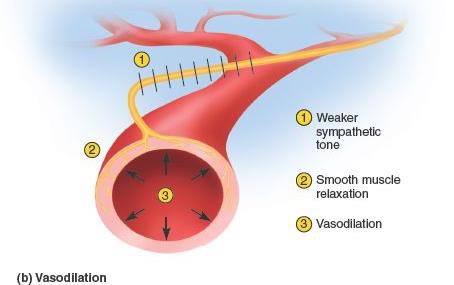
Skeletal Muscle Blood Vessels: ACh causes vasodilation, increasing blood flow during exercise or stress.
Neurotransmitter Release Mechanism
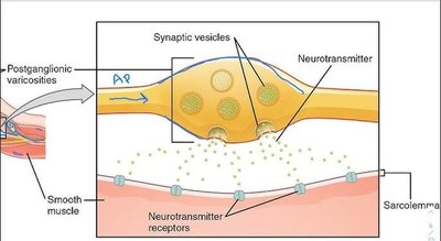
Postganglionic sympathetic neurons have telodendria with swellings called varicosities, which are packed with neurotransmitter vesicles. These varicosities release neurotransmitters onto target cells, where they bind to specific receptors.
Adrenergic Receptors and Their Effects
Types of Adrenergic Receptors
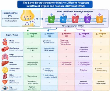
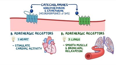
Adrenergic receptors are membrane proteins on target organs that bind norepinephrine and epinephrine. The main types are:
Alpha (α) receptors: α1 and α2
Beta (β) receptors: β1, β2, and β3
The same neurotransmitter can produce different effects depending on the receptor subtype and tissue location.
Examples of Receptor-Specific Effects
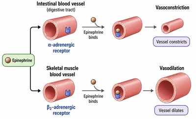
α-adrenergic receptors: Cause vasoconstriction in intestinal blood vessels.
β2-adrenergic receptors: Cause vasodilation in skeletal muscle blood vessels.
Adrenergic Agonists and Antagonists
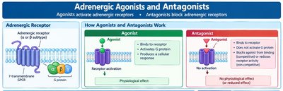
Pharmaceutical agents can act as agonists (activators) or antagonists (blockers) of adrenergic receptors, influencing physiological responses for therapeutic purposes.
Drug | Main Effects / Uses | Receptor |
|---|---|---|
Phenylephrine (α agonist) | Vasoconstriction, ↑ BP; nasal decongestion | α1 |
Prazosin (α antagonist) | Vasodilation, ↓ BP; used for hypertension, BPH | α1 |

Clinical Application: Beta-2 Agonists in Asthma
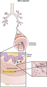
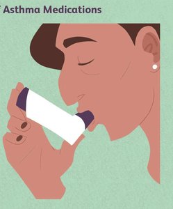
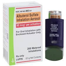
Beta-2 agonists (e.g., albuterol) are used to treat asthma by causing bronchodilation in the lungs, relaxing smooth muscle and improving airflow.
Clinical Application: Epinephrine with Local Anesthetics
Epinephrine is often mixed with local anesthetics to cause vasoconstriction, which confines the anesthetic to the site of administration. This prolongs the drug's local effect, reduces systemic absorption, and minimizes bleeding in the area.
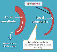

Example: Dental procedures often use local anesthetics with epinephrine for these reasons.
