 Back
BackThe Axial and Appendicular Skeleton: Structure, Function, and Clinical Relevance
Study Guide - Smart Notes
 Tailored notes based on your materials, expanded with key definitions, examples, and context.
The Human Skeleton: Overview
Divisions and Functions
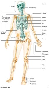
The human skeleton is a framework of bones, cartilages, joints, and ligaments that provides support, protection, and movement. It is divided into two major divisions: the axial skeleton and the appendicular skeleton.
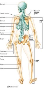
Axial skeleton: Forms the longitudinal axis of the body and includes the skull, vertebral column, and thoracic cage. It supports the head, neck, and trunk, and protects the brain, spinal cord, and thoracic organs.
Appendicular skeleton: Comprises the bones of the limbs and girdles, facilitating movement and manipulation of the environment.
The Skull
Structure and Divisions
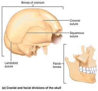
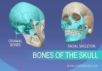
The skull is a complex structure formed by two sets of bones: cranial bones and facial bones. Most skull bones are flat and joined by immovable joints called sutures.
Cranial bones (cranium): Enclose and protect the brain, provide attachment sites for head and neck muscles.
Facial bones: Form the framework of the face, house cavities for special sense organs, provide openings for air and food, secure teeth, and anchor facial muscles.
Major Functions: Cranium vs. Facial Skeleton
Cranium: Protection of the brain and attachment for muscles.
Facial skeleton: Framework for the face, cavities for sensory organs, and muscle attachment for facial expression.
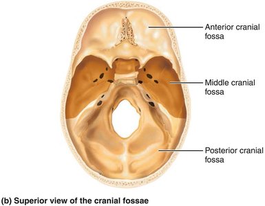
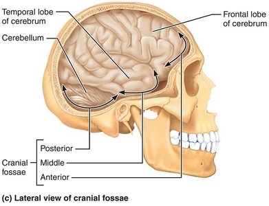
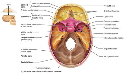
Skull Geography
The cranium is divided into a vault (calvaria) and a base. The base is internally divided into three cranial fossae: anterior, middle, and posterior, which house different parts of the brain.
Other Skull Cavities and Openings
Orbits: House the eyeballs.
Nasal cavity: Passage for air and olfaction.
Sinuses: Air-filled spaces that lighten the skull and enhance voice resonance.
Foramina, canals, fissures: Passageways for nerves and blood vessels.
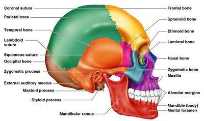
Major Cranial Bones and Markings
Frontal bone: Forms the forehead and superior orbits; contains the frontal sinuses.
Parietal bones: Form most of the superior and lateral aspects of the skull; joined by coronal, sagittal, lambdoid, and squamous sutures.
Occipital bone: Forms the posterior skull and cranial base; contains the foramen magnum and occipital condyles.
Temporal bones: Form the inferolateral skull and part of the cranial base; house structures of the ear and provide muscle attachment points.
Sphenoid bone: Keystone bone that articulates with all other cranial bones; contains the sella turcica and sphenoidal sinuses.
Ethmoid bone: Deepest skull bone; forms part of the nasal septum and medial orbit walls; contains ethmoidal air cells.
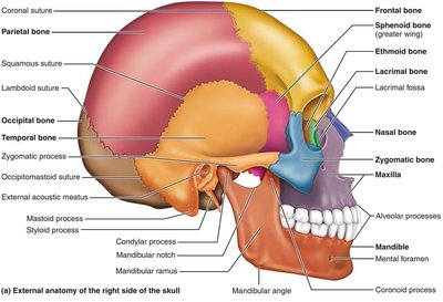
Facial Bones
Mandible: Lower jawbone; largest and strongest facial bone.
Maxillae: Form the upper jaw and central facial skeleton; contain maxillary sinuses.
Zygomatic bones: Form the cheekbones and part of the orbit.
Nasal bones: Form the bridge of the nose.
Lacrimal bones: Form part of the medial orbit wall; house the lacrimal sac.
Palatine bones: Form the posterior hard palate and part of the nasal cavity and orbit.
Vomer: Forms part of the nasal septum.
Inferior nasal conchae: Form part of the lateral walls of the nasal cavity.
Bony Boundaries of the Orbits and Nasal Cavity
Orbits: Formed by the frontal, sphenoid, zygomatic, maxilla, palatine, lacrimal, and ethmoid bones.
Nasal cavity: Formed by the nasal, frontal, ethmoid, sphenoid, maxilla, palatine, vomer, and inferior nasal conchae.
Paranasal Sinuses
Frontal, ethmoidal, sphenoidal, and maxillary sinuses: Air-filled spaces that lighten the skull and enhance voice resonance.
The Fetal Skull
Infant skull has more bones than the adult skull; bones are connected by fontanelles (fibrous membranes) that allow for brain growth and ease birth.
Fontanelles ossify within 24 months after birth.
The Vertebral Column
General Structure and Regions
The vertebral column, or spine, is a flexible, curved structure composed of 26 vertebrae in adults, divided into five regions:
Cervical (7): Neck region.
Thoracic (12): Upper back, articulates with ribs.
Lumbar (5): Lower back, supports the most weight.
Sacrum (1): Formed from fusion of five vertebrae; articulates with the hip bones.
Coccyx (1): Formed from fusion of three to five small vertebrae; the tailbone.
Curvatures and Functions
Normal curvatures: Cervical and lumbar (convex anteriorly), thoracic and sacral (concave anteriorly).
Functions: Transmit weight, protect the spinal cord, provide attachment points for ribs and muscles, and allow flexibility and movement.
Spinal Curvatures and Intervertebral Discs
Spinal curvatures: Increase resilience and flexibility, help maintain balance and posture.
Intervertebral discs: Cushion vertebrae, absorb shock, and allow bending and twisting.
Abnormal Spinal Curvatures
Scoliosis: Abnormal lateral curvature, often in the thoracic region.
Kyphosis: Exaggerated thoracic curvature (hunchback).
Lordosis: Exaggerated lumbar curvature (swayback).
Structure of a Typical Vertebra
Body: Main weight-bearing region.
Vertebral arch: Formed by pedicles and laminae.
Processes: Spinous (posterior), transverse (lateral), superior and inferior articular processes (for articulation with adjacent vertebrae).
Regional Features
Cervical vertebrae: Smallest, have transverse foramina, bifid spinous processes (C2–C6).
Thoracic vertebrae: Heart-shaped body, long spinous processes, facets for rib articulation.
Lumbar vertebrae: Largest, kidney-shaped body, short and blunt spinous processes.
The Thoracic Cage
Structure and Function
Composed of: Sternum, ribs, and thoracic vertebrae.
Functions: Protects heart and lungs, supports shoulder girdle, aids in respiration.
Bones of the Thoracic Cage
Sternum: Manubrium (superior), body (middle), xiphoid process (inferior).
Ribs: 12 pairs—true ribs (1–7, direct attachment to sternum), false ribs (8–12, indirect or no attachment), floating ribs (11–12, no attachment).
The Appendicular Skeleton
Pectoral (Shoulder) Girdle
Clavicle: Collarbone; articulates with sternum and scapula.
Scapula: Shoulder blade; triangular bone with three borders and a prominent spine.
Upper Limb
Humerus: Arm bone; articulates with scapula, radius, and ulna.
Ulna: Medial forearm bone; forms elbow joint with humerus.
Radius: Lateral forearm bone; major contributor to wrist joint.
Hand: Carpals (wrist, 8 bones), metacarpals (palm, 5 bones), phalanges (fingers, 14 bones).
Pelvic Girdle
Hip bones: Each formed by fusion of ilium, ischium, and pubis; articulates at acetabulum.
Functions: Supports upper body weight, protects pelvic organs.
Lower Limb
Femur: Thigh bone; largest and strongest bone in the body.
Patella: Kneecap; protects knee joint.
Tibia: Medial leg bone; bears weight.
Fibula: Lateral leg bone; does not bear weight.
Foot: Tarsals (ankle, 7 bones), metatarsals (sole, 5 bones), phalanges (toes, 14 bones).
Arches of the Foot
Medial and lateral longitudinal arches, transverse arch: Support body weight and provide springiness to the step.
Joints (Articulations)
Classification
Structural: Fibrous (immovable), cartilaginous (slightly movable), synovial (freely movable, joint cavity present).
Functional: Synarthroses (immovable), amphiarthroses (slightly movable), diarthroses (freely movable).
Synovial Joints
Articulating bones separated by a joint cavity filled with synovial fluid.
Features: Articular cartilage, joint capsule, synovial membrane, ligaments, bursae, and tendon sheaths.
Movement depends on articular surface shape, ligament number/position, and muscle tone.
Inflammatory Conditions
Bursitis: Inflammation of a bursa.
Tendonitis: Inflammation of tendon sheaths.
Arthritis: Over 100 types; osteoarthritis (degenerative), rheumatoid arthritis (autoimmune).
Clinical and Preventive Aspects of Bone Health
Bone Density and Imaging
X-rays, MRI, CT scans: Used to assess bone structure and diagnose fractures or abnormalities.
DEXA scan: Measures bone density and osteoporosis risk.
Hormonal Regulation
Estrogen and testosterone: Promote bone deposition and density.
Parathyroid hormone (PTH): Regulates calcium levels and stimulates bone resorption when needed.
Exercise and Prevention
Weight-bearing and resistance exercises increase bone density and reduce fracture risk.
Prevention includes adequate calcium/vitamin D, exercise, and avoiding smoking/excess alcohol.
Rehabilitation
Physical and occupational therapy are crucial for recovery after fractures, improving strength, flexibility, and independence.
