 Back
BackThe Axial and Appendicular Skeleton: Structure, Function, and Clinical Relevance
Study Guide - Smart Notes
 Tailored notes based on your materials, expanded with key definitions, examples, and context.
The Human Skeleton: Overview
Divisions and Functions
The human skeleton is a framework of bones, cartilages, joints, and ligaments that provides support, protection, and movement. It is divided into two major divisions: the axial skeleton and the appendicular skeleton.
Axial skeleton: Forms the longitudinal axis of the body, supporting the head, neck, and trunk, and protecting the brain, spinal cord, and thoracic organs.
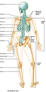
Appendicular skeleton: Composed of the limbs and girdles, facilitating movement and interaction with the environment.
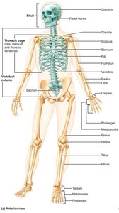
The Axial Skeleton
Main Components
Skull
Vertebral column
Thoracic cage
The axial skeleton consists of 80 bones and serves to protect vital organs and provide structural support.
The Skull
Structure and Divisions
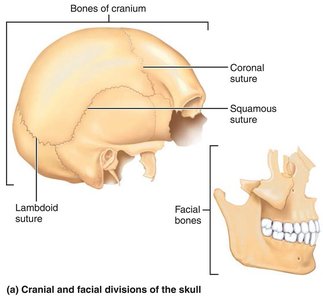
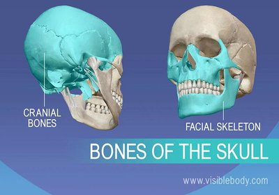
The skull is formed by two sets of bones: cranial bones (cranium) and facial bones. Most skull bones are flat and joined by immovable joints called sutures.
Cranial bones: Enclose and protect the brain, provide attachment sites for head and neck muscles.
Facial bones: Form the framework of the face, contain cavities for sensory organs, provide openings for air and food, secure teeth, and anchor facial muscles.
Major Functions: Cranium vs. Facial Skeleton
Cranium: Protection of the brain and attachment for muscles.
Facial skeleton: Framework for the face, cavities for sensory organs, and muscle attachment for facial expression.
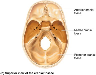
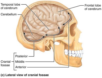
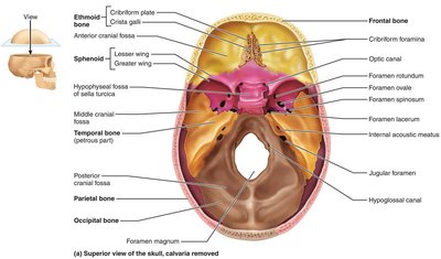
Skull Geography
Cranial vault (calvaria): Forms the superior, lateral, and posterior portions of the skull.
Cranial base: Forms the inferior aspect, divided into anterior, middle, and posterior cranial fossae.
Cranial cavity: Encloses the brain.
Other Cavities and Openings
Orbits: House the eyeballs.
Nasal cavity: Passage for air and olfaction.
Sinuses: Air-filled spaces that lighten the skull and enhance voice resonance.
Foramina, canals, fissures: Passageways for nerves and blood vessels.
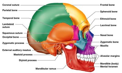
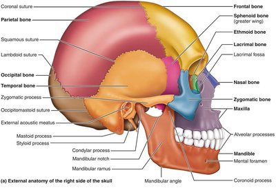
Major Skull Bones and Markings
Frontal bone: Forms the forehead and superior orbits; contains the glabella and frontal sinuses.
Parietal bones: Form the superior and lateral aspects; joined by coronal, sagittal, lambdoid, and squamous sutures.
Occipital bone: Forms the posterior skull; contains the foramen magnum, occipital condyles, and nuchal lines.
Temporal bones: Form the inferolateral skull; include the zygomatic process, external acoustic meatus, mastoid and styloid processes.
Sphenoid bone: Keystone bone that articulates with all cranial bones; contains the sella turcica and sphenoidal sinuses.
Ethmoid bone: Deepest skull bone; forms part of the nasal septum and orbits; contains cribriform plates and ethmoidal air cells.
Facial Bones
Mandible: Lower jawbone, largest and strongest facial bone; contains alveolar processes for teeth.
Maxillae: Upper jaw, forms central facial skeleton and hard palate; contains maxillary sinuses.
Zygomatic bones: Form cheekbones and part of the orbit.
Nasal bones: Form the bridge of the nose.
Lacrimal bones: Form part of the medial orbit wall; contain lacrimal fossa for tear drainage.
Palatine bones: Form posterior hard palate and part of the nasal cavity and orbit.
Vomer: Forms part of the nasal septum.
Inferior nasal conchae: Form part of the lateral walls of the nasal cavity.
Bony Boundaries of Orbits and Nasal Cavity
Orbits: Formed by frontal, sphenoid, zygomatic, maxilla, palatine, lacrimal, and ethmoid bones.
Nasal cavity: Roof (nasal, frontal, ethmoid, sphenoid), floor (maxilla, palatine), lateral walls (maxilla, ethmoid, inferior nasal conchae, palatine), septum (ethmoid, vomer).
Paranasal Sinuses
Frontal, ethmoidal, sphenoidal, maxillary sinuses: Lighten the skull and enhance voice resonance.
The Fetal Skull
Infant skull has more bones than adult skull; bones are connected by fontanelles (fibrous membranes) that allow for brain growth and ease birth.
Fontanelles ossify within 24 months after birth.
The Vertebral Column
Structure and Regions
The vertebral column, or spine, is a flexible, curved structure composed of 26 vertebrae in adults, divided into five regions:
Cervical (7): Neck region
Thoracic (12): Upper back
Lumbar (5): Lower back
Sacrum (1): Fusion of 5 sacral vertebrae
Coccyx (1): Fusion of 3-5 coccygeal vertebrae
Vertebrae are separated by intervertebral discs that provide shock absorption and flexibility.
Curvatures
Cervical and lumbar: Convex anteriorly
Thoracic and sacral: Concave anteriorly
Curvatures increase resilience and flexibility, helping maintain balance and absorb shock.
Abnormal Spinal Curvatures
Scoliosis: Lateral curvature, often in thoracic region
Kyphosis: Exaggerated thoracic curvature (hunchback)
Lordosis: Exaggerated lumbar curvature (swayback)
Structure of a Typical Vertebra
Body: Weight-bearing region
Vertebral arch: Formed by pedicles and laminae
Processes: Spinous (posterior), transverse (lateral), superior and inferior articular processes
Regional Features
Cervical: Smallest, have transverse foramina, bifid spinous processes (C2–C6)
Thoracic: Heart-shaped body, long spinous processes, facets for ribs
Lumbar: Largest, kidney-shaped body, short blunt spinous processes
The Thoracic Cage
Components and Functions
Sternum: Manubrium, body, xiphoid process
Ribs: 12 pairs (true ribs 1–7, false ribs 8–12, floating ribs 11–12)
Thoracic vertebrae: Posterior anchor
The thoracic cage protects the heart and lungs, supports the shoulder girdle, and aids in respiration.
True vs. False Ribs
True ribs (1–7): Attach directly to sternum via costal cartilage
False ribs (8–12): Attach indirectly or not at all (floating ribs 11–12)
The Appendicular Skeleton
Pectoral Girdle and Upper Limb
Pectoral girdle: Clavicle and scapula; allows free movement of the upper limb
Humerus: Arm bone, articulates with scapula, radius, and ulna
Ulna and radius: Forearm bones; ulna forms elbow joint, radius contributes to wrist joint
Hand: Carpals (wrist, 8 bones), metacarpals (palm, 5 bones), phalanges (fingers, 14 bones)
Pelvic Girdle and Lower Limb
Pelvic girdle: Hip bones (ilium, ischium, pubis); supports upper body weight, protects organs
Femur: Thigh bone, largest and strongest bone
Patella: Kneecap, protects knee joint
Tibia and fibula: Leg bones; tibia bears weight, fibula stabilizes ankle
Foot: Tarsals (ankle, 7 bones), metatarsals (sole, 5 bones), phalanges (toes, 14 bones)
Pelvic Differences
Female pelvis: Wider, shallower, lighter, adapted for childbirth
Male pelvis: Narrower, deeper, heavier
Joints (Articulations)
Classification
Structural: Fibrous (immovable), cartilaginous (slightly movable), synovial (freely movable, joint cavity present)
Functional: Synarthroses (immovable), amphiarthroses (slightly movable), diarthroses (freely movable)
Synovial Joints
Articular cartilage covers bone ends
Joint cavity filled with synovial fluid
Enclosed by a fibrous capsule and synovial membrane
Reinforced by ligaments and muscle tone
Associated structures: bursae (fluid-filled sacs), tendon sheaths
Types of Synovial Joints (by shape)
Plane, hinge, pivot, condyloid, saddle, ball-and-socket
Inflammatory Joint Conditions
Bursitis: Inflammation of a bursa
Tendonitis: Inflammation of tendon sheaths
Arthritis: Over 100 types; osteoarthritis (degenerative), rheumatoid arthritis (autoimmune)
Clinical and Preventive Aspects of Bone Health
Bone Density and Imaging
X-rays, MRI, CT scans: Used to assess bone structure and diagnose fractures or abnormalities
DEXA scan: Measures bone density, screens for osteoporosis
Hormonal Regulation
Estrogen and testosterone: Promote bone deposition and density
Parathyroid hormone (PTH): Regulates calcium levels, stimulates bone resorption
Exercise and Bone Health
Weight-bearing and resistance exercises increase bone density and reduce fracture risk
Flexibility exercises improve range of motion and reduce injury risk
Rehabilitation and Therapy
Physical and occupational therapy aid recovery after fractures
Rehabilitation restores function and mobility
Prevention Strategies
Adequate calcium and vitamin D intake
Regular exercise
Avoiding smoking and excessive alcohol
Risk Factors for Osteoporosis
Postmenopausal women (decreased estrogen)
Age, genetics, lifestyle factors
Summary Table: Major Bones of the Axial Skeleton
Region | Main Bones | Key Functions |
|---|---|---|
Skull | Frontal, parietal, occipital, temporal, sphenoid, ethmoid, mandible, maxilla, zygomatic, nasal, lacrimal, palatine, vomer, inferior nasal conchae | Protects brain, forms face, houses sensory organs |
Vertebral Column | Cervical (7), thoracic (12), lumbar (5), sacrum, coccyx | Supports trunk, protects spinal cord, allows movement |
Thoracic Cage | Sternum, ribs (true, false, floating), thoracic vertebrae | Protects heart and lungs, supports shoulder girdle |
Additional info: This guide integrates foundational anatomy, clinical relevance, and preventive strategies for bone health, suitable for ANP college-level study.
