 Back
BackThe Axial Skeleton: Structure and Function of the Skull and Vertebral Column
Study Guide - Smart Notes
 Tailored notes based on your materials, expanded with key definitions, examples, and context.
The Axial Skeleton
Overview of the Axial Skeleton
The axial skeleton forms the central axis of the human body and is essential for protection, support, and movement. It consists of the skull, vertebral column, and thoracic cage. This section focuses on the structure and function of the skull and vertebral column, highlighting their anatomical features and clinical significance.
The Skull
General Structure and Function
The skull is composed of two main sets of bones: cranial bones and facial bones. Cranial bones enclose the brain within the cranial cavity, while facial bones form the framework of the face, house special sense organs, and provide openings for air and food passage. Most skull bones are flat and joined by immovable joints called sutures.
Cranial bones: Protect the brain and form the cranial cavity.
Facial bones: Support the face, secure teeth, and form cavities for the senses.
Sutures: Immovable joints connecting most skull bones (except the mandible).
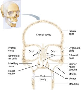
Skull Cavities and Openings
The skull contains several important cavities and numerous openings that serve as passageways for nerves and blood vessels.
Cranial cavities: Anterior, middle, and posterior cranial fossae house different parts of the brain.
Other cavities: Middle and internal ear cavities, nasal cavity, and orbits (eye sockets).
Openings: Foramina, canals, and fissures provide routes for the spinal cord, blood vessels, and cranial nerves.
Cranial Bones
There are eight cranial bones:
Frontal bone
Parietal bones (left and right)
Occipital bone
Temporal bones (left and right)
Sphenoid bone
Ethmoid bone
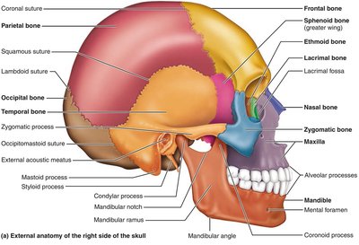
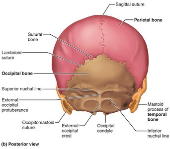
Key Features of Selected Cranial Bones
Frontal bone: Forms the forehead and superior part of the orbits; contains the supraorbital margin and foramen.
Parietal bones: Joined by four major sutures: coronal, sagittal, lambdoid, and squamous.
Occipital bone: Contains the foramen magnum, occipital condyles, and external occipital protuberance.
Temporal bones: Divided into squamous, tympanic, and petrous parts; features include the zygomatic process, external acoustic meatus, mastoid process, and styloid process.
Sphenoid bone: Keystone bone that articulates with all other cranial bones; features include the sella turcica, optic canals, and pterygoid processes.
Ethmoid bone: Contains the crista galli, cribriform plates, perpendicular plate, and superior/middle nasal conchae.
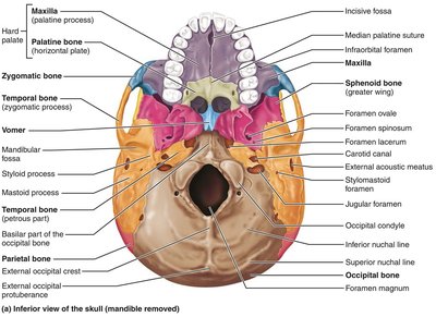



Facial Bones
The facial skeleton consists of 14 bones that form the structure of the face and support the teeth and special sense organs.
Mandible (lower jaw)
Maxillary bones (upper jaw, paired)
Zygomatic bones (cheekbones, paired)
Nasal bones (paired)
Lacrimal bones (paired)
Palatine bones (paired)
Vomer
Inferior nasal conchae (paired)

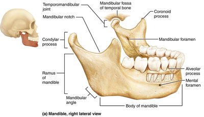

Key Features of Selected Facial Bones
Mandible: Largest and strongest facial bone; features include the mandibular angle, condylar process, coronoid process, alveolar process, mandibular foramen, and mental foramen.
Maxillary bones: Form the upper jaw and part of the hard palate; contain alveolar processes and infraorbital foramen.
Zygomatic bones: Form the prominences of the cheeks and part of the lateral wall of the orbits.
Nasal bones: Form the bridge of the nose.
Lacrimal bones: Small bones forming part of the medial wall of each orbit.
Palatine bones: L-shaped bones forming the posterior part of the hard palate and part of the nasal cavity.
Vomer: Forms part of the nasal septum.
Inferior nasal conchae: Thin, curved bones in the nasal cavity.
Other Skull Structures
Hyoid bone: The only bone that does not articulate directly with another bone; serves as a movable base for the tongue and attachment site for muscles involved in swallowing and speech.
Orbits: Cavities that encase the eyes and lacrimal glands, formed by parts of seven bones.
Paranasal sinuses: Air-filled spaces in five skull bones (frontal, sphenoid, ethmoid, and paired maxillary bones) that warm and humidify air, lighten the skull, and enhance voice resonance.
The Vertebral Column
General Characteristics
The vertebral column, or spine, extends from the skull to the pelvis and is composed of 26 irregular bones called vertebrae. It transmits the weight of the trunk to the lower limbs, surrounds and protects the spinal cord, and provides attachment points for ribs and muscles. The vertebral column has four normal curves that increase its strength and flexibility.
Regions: Cervical, thoracic, lumbar, sacral, and coccygeal.
Curves: Cervical and lumbar (anteriorly convex); thoracic and sacral (anteriorly concave).
Functions of curves: Increase strength, maintain balance, absorb shock, and protect from fractures.
Vertebral Column Components
Ligaments: Support the vertebral column and prevent excessive movement (anterior/posterior longitudinal ligaments, ligamentum flavum).
Intervertebral discs: Cushion-like pads composed of a gelatinous nucleus pulposus and a collagen/fibrocartilage anulus fibrosus.
Clinical Note: Herniated (prolapsed) discs occur when the anulus fibrosus ruptures, allowing the nucleus pulposus to protrude and compress nerves, causing pain or numbness.
Regional Vertebral Characteristics
Cervical vertebrae (C1–C7): Small, oval bodies; bifid spinous processes; transverse foramina; C1 (atlas) and C2 (axis) specialized for head movement.
Thoracic vertebrae (T1–T12): Heart-shaped bodies; long, downward-pointing spinous processes; facets for rib articulation.
Lumbar vertebrae (L1–L5): Massive, kidney-shaped bodies; short, blunt spinous processes; triangular vertebral foramen.
Sacrum: Five fused vertebrae forming the posterior wall of the pelvis; features include the sacral promontory, alae, and sacral foramina.
Coccyx: Three to five fused vertebrae forming the tailbone.
Characteristic | Cervical (C3–C7) | Thoracic | Lumbar |
|---|---|---|---|
Body | Small, oval, wide side to side | Larger than cervical; heart-shaped; bears two costal facets | Massive; kidney-shaped |
Spinous process | Short; bifid (except C7); projects directly posterior | Long; sharp; projects inferiorly | Short; blunt; rectangular; projects directly posteriorly |
Vertebral foramen | Triangular, large | Circular | Triangular |
Transverse processes | Contain foramina | Bear facets for ribs (except T11 and T12) | Thin and tapered |
Movements allowed | Flexion, extension, lateral flexion, rotation; greatest range of movement | Rotation; lateral flexion possible but restricted by ribs; flexion and extension limited | Flexion, extension, some lateral flexion; rotation prevented |
Thoracic Cage
Structure and Function
The thoracic cage consists of the thoracic vertebrae, ribs, sternum, and costal cartilages. It protects vital organs, supports the shoulder girdles and upper limbs, and provides attachment sites for muscles.
Sternum: Composed of the manubrium, body, and xiphoid process.
Ribs: 12 pairs; true ribs (1–7), false ribs (8–10), and floating ribs (11–12).
*Additional info: The above notes are expanded with academic context to ensure completeness and clarity for college-level anatomy and physiology students.*
