 Back
BackThe Axial Skeleton: Structure and Function
Study Guide - Smart Notes
 Tailored notes based on your materials, expanded with key definitions, examples, and context.
Chapter 7: The Axial Skeleton
Divisions of the Skeletal System
The human skeleton is divided into two main parts: the axial skeleton and the appendicular skeleton. The axial skeleton forms the central axis of the body and is essential for protection, support, and movement.
Axial Skeleton: Includes the skull, vertebral column (spine), and thoracic cage (ribs and sternum). It supports the head, neck, back, and chest, and protects the brain, spinal cord, and thoracic organs.
Appendicular Skeleton: Composed of the bones of the upper and lower limbs, as well as the girdles (pectoral and pelvic) that attach the limbs to the axial skeleton.
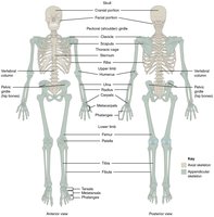
Example: The vertebral column is part of the axial skeleton, while the femur is part of the appendicular skeleton.
The Skull
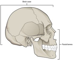
The skull is a complex structure that protects the brain and forms the structure of the face. It consists of two main sets of bones: the brain case (cranial bones) and the facial bones.
Brain Case (Cranial Bones): Surround and protect the brain. Includes the frontal, parietal, temporal, occipital, sphenoid, and ethmoid bones.
Facial Bones: Form the structure of the face, including the upper and lower jaws, nose, and orbits (eye sockets).
Major Suture Lines: Immovable joints between skull bones, such as the coronal, sagittal, squamous, and lambdoid sutures.
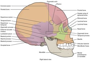
Example: The mandible is the only moveable bone of the skull, forming the lower jaw.
Cranial Fossae and Sinuses
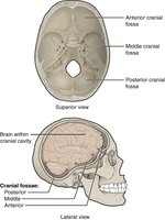
The base of the cranial cavity is divided into three fossae: anterior, middle, and posterior. The paranasal sinuses are air-filled spaces within certain skull bones that reduce skull weight and contribute to voice resonance.
Anterior Cranial Fossa: Supports the frontal lobes of the brain.
Middle Cranial Fossa: Supports the temporal lobes.
Posterior Cranial Fossa: Supports the cerebellum.
Paranasal Sinuses: Frontal, maxillary, sphenoid, and ethmoid sinuses.
Special Features of the Skull
Orbit: Formed by seven bones; houses the eye and associated structures.
Nasal Septum: Formed by the perpendicular plate of the ethmoid bone and the vomer.
Hyoid Bone: Located in the neck, does not articulate with other bones, supports the tongue and larynx.
The Vertebral Column
Regions and Curvatures
The vertebral column, or spine, is composed of 24 vertebrae, the sacrum, and the coccyx. It provides structural support, protects the spinal cord, and allows flexible movement.
Cervical Region: 7 vertebrae (C1–C7)
Thoracic Region: 12 vertebrae (T1–T12)
Lumbar Region: 5 vertebrae (L1–L5)
Sacrum: 5 fused vertebrae
Coccyx: 4 fused vertebrae
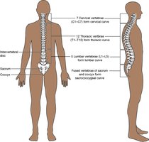
Spinal Curvatures: Primary (thoracic, sacrococcygeal) and secondary (cervical, lumbar) curves help balance the body and absorb shock.
Abnormal Spinal Curvatures
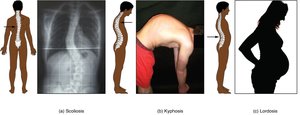
Scoliosis: Lateral curvature of the spine.
Kyphosis: Excessive thoracic curvature (hunchback).
Lordosis: Excessive lumbar curvature (swayback).
Structure of a Typical Vertebra
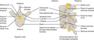
A typical vertebra consists of a vertebral body, vertebral arch (formed by pedicles and laminae), and several processes for muscle and ligament attachment.
Vertebral Foramen: Passage for the spinal cord.
Intervertebral Foramen: Opening for spinal nerves between adjacent vertebrae.
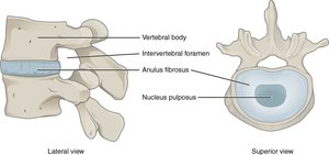
Intervertebral Discs: Pads of fibrocartilage (anulus fibrosus and nucleus pulposus) that cushion and allow movement between vertebrae.
Regional Vertebral Differences
Cervical Vertebrae: Small body, bifid spinous process, transverse foramen. Atlas (C1) and axis (C2) are specialized for head movement.
Thoracic Vertebrae: Long, downward spinous process, facets for rib articulation.
Lumbar Vertebrae: Large, thick body, short spinous process for weight bearing.
Sacrum and Coccyx: Fused vertebrae forming the posterior pelvic wall.
Ligaments of the Vertebral Column
Anterior Longitudinal Ligament: Runs along the anterior surface of vertebral bodies.
Supraspinous Ligament: Connects the tips of the spinous processes.
Nuchal Ligament: Enlarged portion in the neck, supports the head.
The Thoracic Cage
Components and Structure
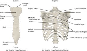
The thoracic cage protects vital organs and supports the upper body. It consists of the sternum, ribs, and thoracic vertebrae.
Sternum: Composed of the manubrium, body, and xiphoid process.
Ribs: 12 pairs, classified as true ribs (1–7), false ribs (8–12), and floating ribs (11–12).
Costal Cartilages: Connect ribs to the sternum.
Embryonic Development of the Axial Skeleton
During embryonic development, bones of the skull and axial skeleton form through two processes: intramembranous and endochondral ossification.
Fontanelles: Soft spots in the newborn skull where bones have not yet fused, allowing for brain growth.
Vertebral Column and Thoracic Cage: Develop from mesenchymal cells that differentiate into cartilage and then bone.
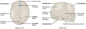
Additional info: Intramembranous ossification forms most cranial bones, while endochondral ossification forms the base of the skull and most of the vertebral column and ribs.
