 Back
BackThe Axial Skeleton: Structure and Function
Study Guide - Smart Notes
 Tailored notes based on your materials, expanded with key definitions, examples, and context.
The Axial Skeleton
Introduction
The axial skeleton forms the central axis of the human body, providing support and protection for the brain, spinal cord, heart, and lungs. It serves as an attachment site for muscles that move the head, neck, and back, as well as those that act across the shoulder and hip joints. The axial skeleton consists of the skull, vertebral column, and thoracic cage, totaling 80 bones in the adult human body.
The Skull
Major Divisions of the Skull
The skull is divided into two main regions: the brain case (neurocranium) and the facial bones (viscerocranium). The brain case surrounds and protects the brain, while the facial bones support the structures of the face, form the nasal cavity, enclose the eyeballs, and support the teeth.
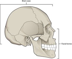
Brain Case: Composed of 8 bones, including paired parietal and temporal bones, and unpaired frontal, occipital, sphenoid, and ethmoid bones.
Facial Bones: Composed of 14 bones, including paired maxillae, zygomatic, nasal, lacrimal, palatine, and inferior nasal conchae, and unpaired mandible and vomer.
Bones of the Brain Case
Frontal Bone: Forms the forehead and the roof of the orbit.
Parietal Bones: Form the upper lateral sides of the skull.
Occipital Bone: Forms the posterior skull and base, containing the foramen magnum for the spinal cord.
Temporal Bones: Form the lower lateral sides of the skull, containing the mastoid and styloid processes, and the external acoustic meatus.
Sphenoid Bone: Keystone bone of the skull, articulating with nearly all other skull bones; contains the sella turcica for the pituitary gland.
Ethmoid Bone: Forms the roof and lateral walls of the nasal cavity and part of the orbit; contains the crista galli and cribriform plate for olfactory nerves.
Facial Bones
The facial bones provide the bony framework for the face, support the teeth, and form the nasal and orbital cavities.
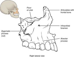
Maxilla: Forms the upper jaw, hard palate, and part of the orbit. Contains the infraorbital foramen for nerves and vessels.
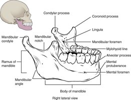
Mandible: The only moveable bone of the skull, forming the lower jaw. Features include the mandibular condyle, coronoid process, mental foramen, and alveolar process.
Zygomatic Bones: Form the cheekbones and part of the orbit.
Nasal Bones: Form the bridge of the nose.
Lacrimal Bones: Small bones forming part of the medial orbit wall.
Palatine Bones: Form the posterior part of the hard palate.
Vomer: Forms the inferior part of the nasal septum.
Sutures of the Skull
Sutures are immobile fibrous joints that tightly unite the bones of the skull. Major sutures include the coronal, sagittal, lambdoid, and squamous sutures. These interlocking joints provide strength and allow for growth during development.
Fetal Skull and Fontanelles
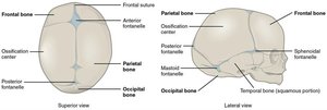
In the fetal skull, bones are separated by large areas of dense connective tissue called fontanelles. These "soft spots" allow for deformation during birth and rapid brain growth after birth. The major fontanelles are the anterior, posterior, mastoid, and sphenoidal fontanelles. The skull bones fuse at the sutures as the child grows.
The Vertebral Column
Regions of the Vertebral Column
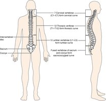
The vertebral column is divided into five regions: cervical (7 vertebrae), thoracic (12 vertebrae), lumbar (5 vertebrae), sacrum (5 fused vertebrae), and coccyx (4 fused vertebrae). It supports the head, protects the spinal cord, and provides attachment points for ribs and muscles.
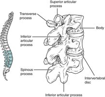
General Structure of a Vertebra
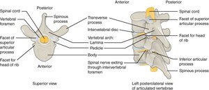
All vertebrae share a common structure: a body (anterior, weight-bearing), a vertebral arch (posterior), and seven processes (spinous, transverse, and articular). The vertebral foramen houses the spinal cord, and intervertebral discs separate adjacent vertebral bodies.
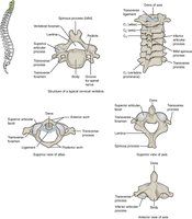
Cervical Vertebrae
Cervical vertebrae are characterized by small bodies, bifid spinous processes (except C7), and transverse foramina for vertebral arteries. The atlas (C1) supports the skull, and the axis (C2) provides a pivot for head rotation.
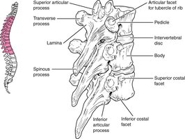
Thoracic Vertebrae
Thoracic vertebrae have larger bodies, long downward-pointing spinous processes, and facets for rib articulation. The orientation of articular processes allows for rotation but limits flexion and extension.
Lumbar Vertebrae
Lumbar vertebrae have the largest bodies to support body weight, short and blunt spinous processes, and large articular processes. They allow for flexion and extension but limit rotation.
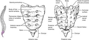
Sacrum and Coccyx
The sacrum is a triangular bone formed by the fusion of five sacral vertebrae, providing a strong foundation for the pelvic girdle. The coccyx, or tailbone, is formed by the fusion of four small coccygeal vertebrae and serves as an attachment site for ligaments and muscles.
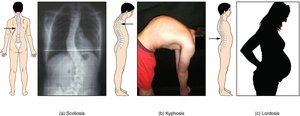
Abnormal Curvatures of the Vertebral Column
Abnormal spinal curvatures include:
Kyphosis: Excessive posterior curvature of the thoracic region (humpback).
Lordosis: Excessive anterior curvature of the lumbar region (swayback).
Scoliosis: Abnormal lateral curvature, often developing during adolescence.
The Thoracic Cage
Structure and Function
The thoracic cage protects the heart and lungs and provides attachment points for muscles involved in respiration and upper limb movement. It consists of the sternum, 12 pairs of ribs, and costal cartilages.
Sternum
Manubrium: Superior portion, articulates with clavicles and first ribs.
Body: Central portion, articulates with costal cartilages of ribs 2–7.
Xiphoid Process: Inferior tip, attachment site for muscles.
Jugular (Suprasternal) Notch: Superior border, palpable at the base of the neck.
Clavicular Notch: Lateral to jugular notch, articulates with clavicles.
Sternal Angle: Junction of manubrium and body, landmark for rib counting.
Ribs
True Ribs (1–7): Attach directly to the sternum via costal cartilages.
False Ribs (8–12): Attach indirectly or not at all to the sternum.
Floating Ribs (11–12): Do not attach to the sternum.
Summary Table: Classification of Ribs
Type | Rib Numbers | Attachment to Sternum |
|---|---|---|
True Ribs | 1–7 | Direct |
False Ribs | 8–12 | Indirect or none |
Floating Ribs | 11–12 | None |
Key Terms and Concepts
Foramen Magnum: Large opening in the occipital bone for the spinal cord.
Fontanelle: Soft spot in the fetal skull where bones have not yet fused.
Articular Process: Projection that forms a joint with an adjacent bone.
Costal Cartilage: Hyaline cartilage connecting ribs to the sternum.
Clinical Applications
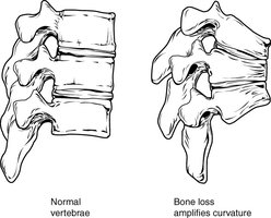
Osteoporosis: Weakening of bone that can lead to kyphosis due to vertebral collapse.
Scoliosis Treatment: May include bracing or surgery in severe cases.
Additional info: The axial skeleton is essential for protecting vital organs and supporting the body’s structure. Understanding its anatomy is foundational for further study in anatomy and physiology, as well as for clinical practice in medicine and allied health fields.
