 Back
Ch 7: The Axial Skeleton: Structure and Function
Study Guide - Smart Notes
 Tailored notes based on your materials, expanded with key definitions, examples, and context.
The Axial Skeleton
Overview of the Axial Skeleton
The axial skeleton forms the central supporting structure of the body and consists of 80 bones, accounting for about 40% of the bones in the human body. It includes the skull, bones associated with the skull, the thoracic cage, and the vertebral column. The axial skeleton provides protection for the brain, spinal cord, and thoracic and abdominal organs, and serves as an attachment site for muscles involved in head, neck, and trunk movements, breathing, and stabilization of the appendicular skeleton.
Skull: 8 cranial bones and 14 facial bones
Associated bones: 6 auditory ossicles and the hyoid bone
Thoracic cage: Sternum and 24 ribs
Vertebral column: 24 vertebrae, sacrum, and coccyx
The Skull
Structure and Function of the Skull
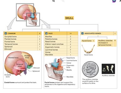
The skull is composed of 22 bones: 8 cranial bones that protect the brain and 14 facial bones that form the structure of the mouth, nose, and orbits. The skull also includes 7 associated bones: the hyoid bone and 6 auditory ossicles. The cranial bones enclose the cranial cavity, providing protection and support for the brain, while the facial bones support the entrances to the digestive and respiratory tracts and provide attachment sites for facial muscles.
Sutures of the Skull
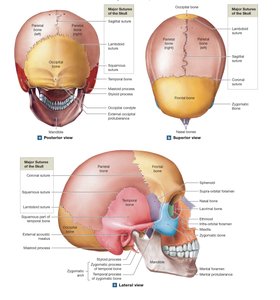
Sutures are immovable joints that connect most of the bones of the adult skull. They are composed of dense fibrous connective tissue. The four major sutures are:
Lambdoid Suture: Connects the occipital bone with the two parietal bones.
Coronal Suture: Attaches the frontal bone to the parietal bones.
Sagittal Suture: Extends from the lambdoid to the coronal suture between the parietal bones.
Squamous Sutures: Join the temporal bone and the parietal bone on each side.
Cranial Bones
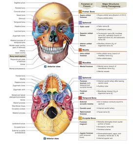
The cranial bones include the occipital, parietal, frontal, temporal, sphenoid, and ethmoid bones. Each bone has distinct features, articulations, and foramina for the passage of nerves and blood vessels.
Occipital Bone
Function: Forms much of the posterior and inferior surfaces of the cranium.
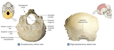
Key Features: External occipital protuberance, external occipital crest, occipital condyles, foramen magnum, hypoglossal canals, nuchal lines.
Articulations: Parietal, temporal, sphenoid bones, and the first cervical vertebra (atlas).
Parietal Bones
Function: Form part of the superior and lateral surfaces of the cranium.
Key Features: Superior and inferior temporal lines, grooves for cranial blood vessels.
Articulations: With each other, occipital, temporal, frontal, and sphenoid bones.
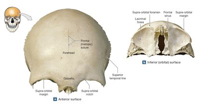
Frontal Bone
Function: Forms the anterior cranium and roof of the orbits; contains frontal sinuses.
Key Features: Forehead, supra-orbital margin, glabella, lacrimal fossa, frontal sinuses.
Articulations: Parietal, sphenoid, ethmoid, nasal, lacrimal, maxillary, and zygomatic bones.
Temporal Bones
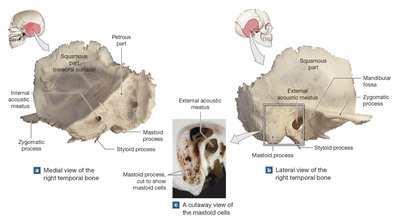
Function: Form part of the lateral walls of the cranium, zygomatic arches, and house the sense organs of the internal ear.
Key Features: Squamous part, zygomatic process, mandibular fossa, mastoid process, styloid process, petrous part, external acoustic meatus.
Articulations: Zygomatic, sphenoid, parietal, occipital bones, and mandible.
Sphenoid
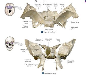
Function: Forms part of the floor of the cranium, unites cranial and facial bones, contains sphenoidal sinuses.
Key Features: Body, sella turcica, hypophyseal fossa, lesser and greater wings, pterygoid processes, optic canals, superior orbital fissure.
Articulations: Frontal, occipital, parietal, temporal, ethmoid, palatine, zygomatic, maxillae, vomer.
Ethmoid
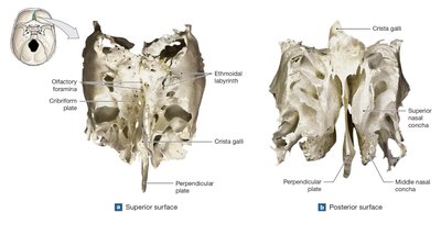
Function: Forms the anteromedial floor of the cranium, roof of the nasal cavity, and part of the nasal septum and medial orbital wall.
Key Features: Cribriform plate, crista galli, ethmoidal labyrinth, perpendicular plate, superior and middle nasal conchae, olfactory foramina.
Articulations: Frontal, sphenoid, maxillae, nasal, lacrimal, palatine, inferior nasal conchae, vomer.
Facial Bones
The facial bones support the entrances to the digestive and respiratory tracts and provide attachment sites for facial muscles. They include the maxillae, palatine bones, nasal bones, vomer, inferior nasal conchae, zygomatic bones, lacrimal bones, and mandible.
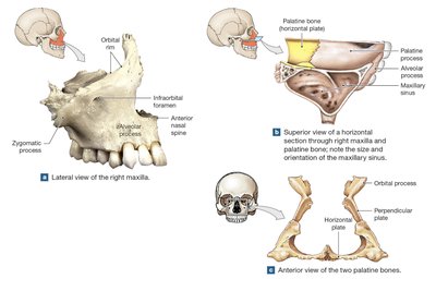
Maxillae
Function: Support upper teeth, form the inferior orbital rims, lateral margins of external nares, upper jaw, and most of the hard palate.
Key Features: Orbital rim, anterior nasal spine, alveolar process, palatine processes, maxillary sinuses, infra-orbital foramen.
Articulations: Frontal, ethmoid, other maxilla, all facial bones except mandible.
Palatine Bones
Function: Form the posterior portions of the hard palate and contribute to the floor of each orbit.
Key Features: Horizontal plate, perpendicular plate, orbital process.
Nasal Bones
Function: Support the superior portions of the bridge of the nose.
Articulations: With each other, ethmoid, frontal, and maxillae.
Vomer
Function: Forms the inferior portion of the bony nasal septum.
Articulations: Maxillae, sphenoid, ethmoid, palatine bones, and cartilaginous nasal septum.
Inferior Nasal Conchae
Function: Slow inhaled air, create turbulence, increase epithelial surface area for warming and humidifying air.
Articulations: Maxillae, ethmoid, palatine, and lacrimal bones.
Zygomatic Bones
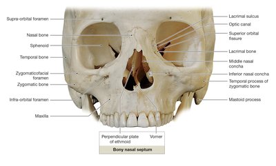
Function: Form the cheekbones and parts of the zygomatic arches, contribute to the rims and lateral walls of the orbits.
Key Features: Temporal process, zygomaticofacial foramen.
Lacrimal Bones
Function: Form parts of the medial walls of the orbits.
Key Features: Lacrimal sulcus, nasolacrimal canal.
Mandible
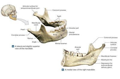
Function: Forms the lower jaw.
Key Features: Body, alveolar part, mental protuberance, ramus, condylar process, coronoid process, mandibular notch, mental foramina, mandibular foramen, mylohyoid line.
Articulations: Mandibular fossae of the temporal bones.
Associated Bones
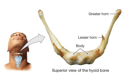
Hyoid Bone: Supports the larynx and is an attachment site for muscles of the larynx, pharynx, and tongue. Connected to the skull by stylohyoid ligaments.
Auditory Ossicles: Three small bones in each middle ear (malleus, incus, stapes) that transfer sound vibrations from the tympanic membrane to the internal ear.
Orbital and Nasal Complexes
Orbital Complex
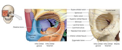
The orbital complex is formed by seven bones and surrounds and protects each eye. The bones include the frontal, maxilla, lacrimal, ethmoid, sphenoid, palatine, and zygomatic bones. Several foramina and fissures allow passage of nerves and blood vessels to the orbit.
Nasal Complex and Paranasal Sinuses
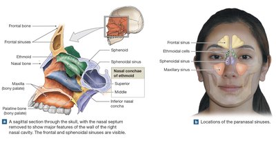
The nasal complex includes bones that enclose the nasal cavities and the paranasal sinuses. The paranasal sinuses are air-filled chambers that lighten the skull, produce mucus, and act as resonating chambers for speech. The bones involved include the frontal, sphenoid, ethmoid, maxillae, and palatine bones.
The Vertebral Column
Regions and General Structure
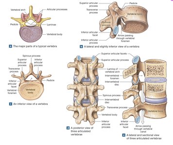
The vertebral column is divided into five regions: cervical (7 vertebrae), thoracic (12 vertebrae), lumbar (5 vertebrae), sacral (5 fused vertebrae), and coccygeal (3–5 fused vertebrae). Each vertebra consists of a vertebral body, vertebral arch (pedicles and laminae), and articular processes. The vertebral column protects the spinal cord, supports the head, and provides attachment sites for muscles and ligaments.
Cervical Vertebrae (C1–C7)
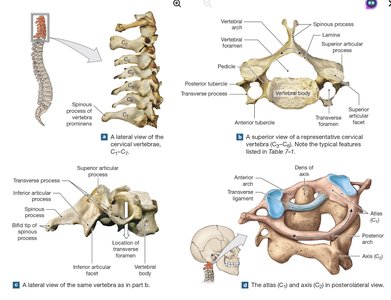
Features: Small body, large vertebral foramen, bifid spinous process (C2–C6), transverse foramina for vertebral arteries and veins.
Atlas (C1): Lacks a body and spinous process; articulates with occipital condyles for nodding motion.
Axis (C2): Has the dens (odontoid process) for rotation of the head.
Vertebra Prominens (C7): Long, prominent spinous process; transition to thoracic region.
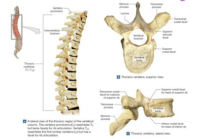
Thoracic Vertebrae (T1–T12)
Features: Heart-shaped body, smaller vertebral foramen, long and slender spinous process, costal facets for rib articulation.
Articulations: Each thoracic vertebra articulates with one or more pairs of ribs.
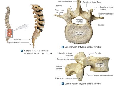
Lumbar Vertebrae (L1–L5)
Features: Largest vertebrae, thick oval body, triangular vertebral foramen, short and stumpy spinous process, no costal facets.
Function: Withstand the most weight and provide attachment for lower back muscles.
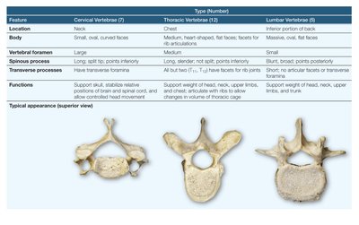
Comparative Table: Cervical, Thoracic, and Lumbar Vertebrae
Feature | Cervical Vertebrae (7) | Thoracic Vertebrae (12) | Lumbar Vertebrae (5) |
|---|---|---|---|
Location | Neck | Chest | Inferior portion of back |
Body | Small, oval, curved faces | Medium, heart-shaped, facets for rib articulation | Massive, oval, flat faces |
Vertebral foramen | Large | Medium | Small |
Spinous process | Long, split tip, points inferiorly | Long, slender, not split, points inferiorly | Short, blunt, points posteriorly |
Transverse processes | Have transverse foramina | All but T11 and T12 have facets for rib articulation | Short, no transverse foramina |
Functions | Support skull, stabilize spinal cord, allow controlled head movement | Support weight of head, neck, upper limbs, articulate with ribs, allow changes in volume of thoracic cage | Support weight of head, neck, upper limbs, trunk, allow flexion and extension |
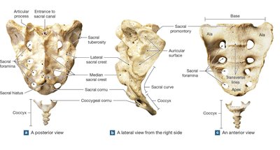
Sacrum and Coccyx
Sacrum: Formed by fusion of five sacral vertebrae; protects pelvic organs and attaches the axial skeleton to the pelvic girdle.
Coccyx: Formed by fusion of 3–5 coccygeal vertebrae; attachment site for ligaments and muscles of the pelvic floor.
The Thoracic Cage
Structure and Function
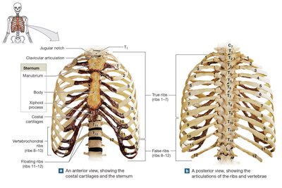
The thoracic cage consists of the thoracic vertebrae, ribs, costal cartilages, and sternum. It protects the heart, lungs, and other thoracic organs, and provides attachment points for muscles involved in breathing, maintaining posture, and moving the pectoral girdles and upper limbs.
True ribs (1–7): Attach directly to the sternum via costal cartilages.
False ribs (8–12): Do not attach directly to the sternum; ribs 8–10 connect via shared cartilage, ribs 11–12 are floating ribs.
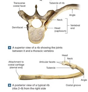
Rib Structure
Head (capitulum): Articulates with vertebral bodies.
Neck and tubercle: Tubercle articulates with the transverse process of thoracic vertebrae.
Body (shaft): Curved, with a costal groove for nerves and blood vessels.
Sternum
Manubrium: Articulates with clavicles and first pair of ribs; contains jugular notch.
Body: Attaches to costal cartilages of ribs 2–7.
Xiphoid process: Smallest part; attachment for diaphragm and rectus abdominis muscle.
Clinical Notes
Temporomandibular Joint Syndrome (TMJ): Involves misalignment and pain in the jaw joint, often due to muscle spasm or stress.
Spinal Curvatures: Kyphosis (excessive thoracic curvature), lordosis (excessive lumbar curvature), and scoliosis (lateral curvature) can result from developmental, muscular, or idiopathic causes.
Summary
The axial skeleton is essential for protecting vital organs, supporting the body, and enabling movement. Understanding the structure and function of its components is fundamental for students of anatomy and physiology.
