Back
BackThe Axial Skeleton: Structure and Landmarks of the Skull
Study Guide - Smart Notes
 Tailored notes based on your materials, expanded with key definitions, examples, and context.
The Axial Skeleton
Overview of the Axial Skeleton
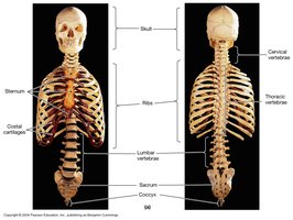
The axial skeleton forms the central axis of the human body and provides support and protection for the brain, spinal cord, and thoracic organs. It consists of the skull, vertebral column, ribs, and sternum.
Skull: Protects the brain and forms the structure of the face.
Vertebral column: Supports the body and protects the spinal cord.
Ribs and sternum: Protect the thoracic organs and assist in respiration.
Sacrum and coccyx: Form the base of the vertebral column.
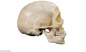
The Skull
General Structure and Classification
The skull is composed of approximately 29 bones, with 11 paired bones. Most joints in the skull are classified as sutural (structural classification) and synarthrosis (functional classification), meaning they are immovable. Exceptions include the temporomandibular joint (TMJ) and the joints of the teeth.
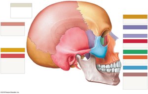
Bones of the Cranium
The cranium consists of 8 bones that form a protective shell around the brain. These bones develop via intramembranous ossification.
Frontal bone
Parietal bones (x2)
Occipital bone
Temporal bones (x2)
Sphenoid bone
Ethmoid bone
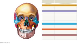
Facial Bones
The facial skeleton is made up of 14 bones that do not directly contact the brain or meninges. These bones provide the structure of the face and house the teeth and nasal cavities.
Maxillae bones (x2)
Zygomatic bones (x2)
Nasal bones (x2)
Inferior nasal conchae bones (x2)
Lacrimal bones (x2)
Vomer bone
Mandible bone
Palatine bones (x2)
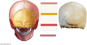
Sutures of the Skull
Major Sutures
Sutures are immovable joints that connect the bones of the skull. The major sutures include:
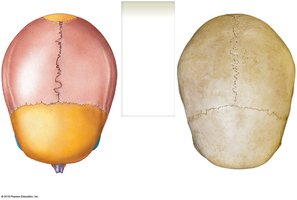
Sagittal suture: Separates the two parietal bones (in the midsagittal plane).
Coronal suture: Separates the frontal bone from the parietal bones.
Lambdoidal (lambdoid) suture: Separates the occipital bone from the parietal bones.
Squamosal suture: Separates the temporal bone from the parietal bone (superior border of the temporal bone).

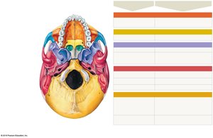
Landmarks of the Cranial Bones
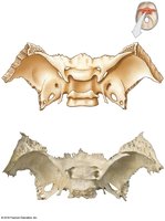
Sphenoid Bone
The sphenoid bone is a complex bone located at the base of the skull. Key landmarks include:
Greater wings
Lesser wings
Optic canal (foramen)
Pterygoid processes
Sella turcica
Superior orbital fissure
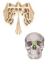
Ethmoid Bone
The ethmoid bone forms part of the nasal cavity and the medial wall of the orbit. Key landmarks include:
Crista galli
Cribriform plate
Perpendicular plate
Middle nasal conchae
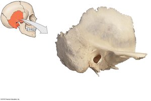
Temporal Bone
The temporal bone houses structures of the ear and forms part of the side and base of the skull. Key landmarks include:
External auditory canal (external acoustic meatus)
Internal auditory canal (internal acoustic meatus)
Mandibular fossa
Mastoid process
Squamous portion (temporal squama)
Styloid process
Zygomatic process
Carotid foramen (canal)
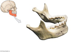
Mandible Bone
The mandible is the lower jawbone and the only movable bone of the skull. Key landmarks include:
Alveolar processes
Mandibular foramen
Mental foramen
Condylar process
Coronoid process
Mandibular notch
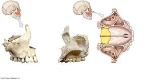
Maxillae Bone
The maxillae form the upper jaw and part of the hard palate. Key landmarks include:
Alveolar processes
Infraorbital foramen
Maxillary sinus
Palatine process
Frontal Bone
Supraorbital foramen: Passage for nerves and blood vessels above the orbit.
Occipital Bone
External occipital protuberance
Foramen magnum
Occipital condyle
Bones and Landmarks of the Nasal Cavity
The nasal cavity is formed by several bones and their landmarks:
Roof: Cribriform plate (ethmoid bone)
Walls (sides): Nasal conchae (ethmoid bone)
Septum: Perpendicular plate (ethmoid bone) and vomer bone
Floor (hard palate): Palatine processes (maxillae bones) and palatine bones
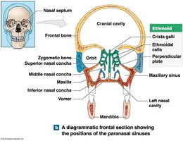
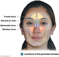
Paranasal Sinuses
Paranasal sinuses are air-filled spaces within certain skull bones. They serve to warm and humidify air before it reaches the lungs and reduce the weight of the skull.
Frontal sinus
Maxillary sinus
Sphenoidal sinus
Ethmoidal air cells
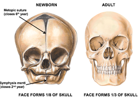
Fetal Skull
Fontanels
Fontanels are regions of fibrous connective tissue between the cranial bones of a developing skull. They provide flexibility for the growing brain and allow distortion of the skull during birth. Most fontanels are fully fused by 12 months of age.
Frontal (anterior) fontanel
Sphenoid (anterolateral) fontanel
Mastoid (posterolateral) fontanel
Occipital (posterior) fontanel
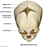
Frontal (Metopic) Suture
The frontal (metopic) suture is present in the fetal skull and typically fuses via synostosis, leaving a remnant in the adult skull. The mental symphysis is another feature that fuses early in development.