Back
BackThe Axial Skeleton: Structure, Disorders, and Clinical Correlations
Study Guide - Smart Notes
 Tailored notes based on your materials, expanded with key definitions, examples, and context.
Chapter 7: The Axial Skeleton
Overview of the Axial Skeleton
The axial skeleton forms the central supporting structure of the human body, consisting of the skull, vertebral column, and thoracic cage. It provides protection for the brain, spinal cord, and thoracic organs, and serves as an attachment point for muscles involved in posture, respiration, and movement.
Major Components: Skull, vertebral column, ribs, sternum, and associated bones (hyoid, auditory ossicles).
Bone Count: The adult axial skeleton contains 80 bones.
Functions: Protection, support, and movement; houses special sense organs.
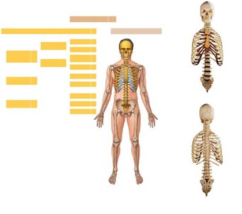
Disorders Affecting the Axial Skeleton
Marfan Syndrome and Gigantism
Several genetic and endocrine disorders can affect the development and structure of the axial skeleton, leading to abnormal growth patterns and skeletal deformities.
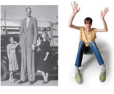
Marfan Syndrome: A connective tissue disorder characterized by rapid elongation of bones, especially in the limbs and fingers, due to abnormal development of the epiphyseal cartilage.
Pituitary Gigantism: Caused by hypersecretion of growth hormone (GH) before and during puberty, resulting in excessive growth of bones.
Pituitary Dwarfism: Hyposecretion of GH before and during development, leading to abnormally short stature.
Cretinism: Congenital hypothyroidism resulting in insufficient thyroxine during early development, causing physical and mental developmental delays.
Myxedema: Hypothyroidism in adults, presenting with symptoms such as lethargy, weight gain, and sluggish mental function.
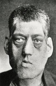
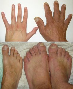
Acromegaly: Hypersecretion of GH after epiphyseal closure, causing thickening of bones in the face, hands, feet, and jaw.
Osteoarthritis
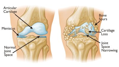
Osteoarthritis is the most common form of arthritis, typically associated with aging or repetitive use of joints. It involves the gradual breakdown of articular cartilage, leading to pain and reduced joint mobility.
Symptoms: Pain, stiffness (especially after inactivity), tenderness, loss of flexibility, grating sensation, bone spurs, and swelling.
Pathology: Loss of cartilage and formation of bone spurs can be observed in affected joints.
Rheumatoid Arthritis
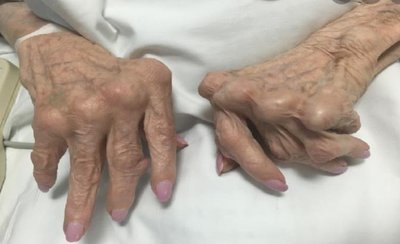
Rheumatoid arthritis (RA) is an autoimmune disorder in which the immune system attacks joint tissues, causing chronic inflammation, pain, and eventual joint damage. Unlike osteoarthritis, RA can affect soft tissues as well as joints.
Mechanism: Immune cells produce proteins such as tumor necrosis factor (TNF), leading to inflammation and tissue destruction.
Symptoms: Pain, swelling, and deformity of joints; systemic effects may occur.
Structural Organization of the Axial Skeleton
Skull and Associated Bones
The skull protects the brain and supports the structures of the face. It is divided into cranial and facial bones, with additional associated bones such as the auditory ossicles and hyoid bone.
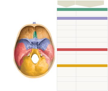
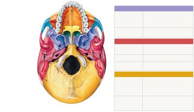
Cranial Bones: Enclose the brain (e.g., frontal, parietal, occipital, temporal, sphenoid, ethmoid).
Facial Bones: Form the structure of the face (e.g., maxilla, zygomatic, mandible, nasal, palatine, vomer, lacrimal, inferior nasal concha).
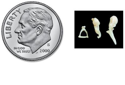
Auditory Ossicles: Three small bones (malleus, incus, stapes) in the middle ear, essential for hearing.
Hyoid Bone: Located in the neck, supports the tongue and larynx.
Major Foramina and Fissures of the Skull
Foramina and fissures in the skull allow the passage of nerves and blood vessels. Key examples include the foramen magnum, optic canal, jugular foramen, and others, each transmitting specific structures.
Foramen Magnum: Passage for the spinal cord, vertebral arteries, and accessory nerve (XI).
Optic Canal: Passage for the optic nerve (II) and ophthalmic artery.
Jugular Foramen: Passage for cranial nerves IX, X, XI, and the internal jugular vein.
The Nasal Complex and Paranasal Sinuses
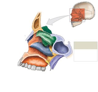
The nasal complex consists of bones and cartilages that form the nasal cavity and paranasal sinuses. These structures warm, moisten, and filter air, and contribute to voice resonance.
Major Bones: Frontal, sphenoid, ethmoid, maxilla, palatine, and nasal bones.
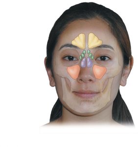
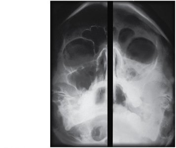
Paranasal Sinuses: Air-filled spaces in the frontal, sphenoid, ethmoid, and maxillary bones; lighten the skull and enhance voice resonance.
Clinical Note: Sinusitis is inflammation of the paranasal sinuses, often causing pain and congestion.
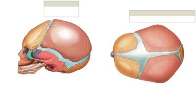
Infant Skull and Fontanelles
Fontanelles are soft spots on an infant's skull where the bones have not yet fused. They allow for flexibility during birth and accommodate rapid brain growth in early life.
Major Fontanelles: Anterior, posterior, sphenoidal, and mastoid fontanelles.
Clinical Importance: Assessment of fontanelles can provide information about hydration and intracranial pressure in infants.
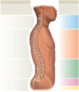
The Vertebral Column
The vertebral column, or spine, is composed of 26 vertebrae in adults, divided into cervical, thoracic, lumbar, sacral, and coccygeal regions. It supports the head, protects the spinal cord, and provides attachment points for ribs and muscles.
Regions: Cervical (7), thoracic (12), lumbar (5), sacrum (1), coccyx (1).
Spinal Curves: Primary curves (thoracic, sacral) develop before birth; secondary curves (cervical, lumbar) develop after birth to support upright posture.
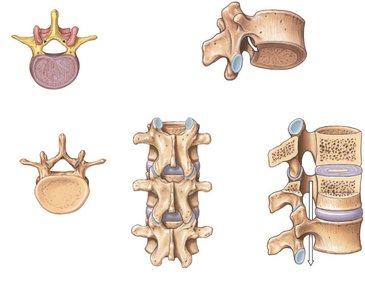
Vertebral Anatomy
A typical vertebra consists of a vertebral body, vertebral arch, and several processes for muscle and ligament attachment. The vertebral foramen forms the vertebral canal, which houses the spinal cord.
Key Structures: Body, pedicle, lamina, spinous process, transverse process, articular processes, vertebral foramen, intervertebral foramen, and intervertebral discs.
Regional Differences in Vertebrae
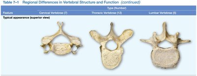
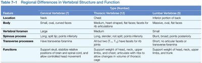
Vertebrae differ in structure and function depending on their region. Cervical vertebrae are small and allow for head movement, thoracic vertebrae articulate with ribs, and lumbar vertebrae are large to support body weight.
Feature | Cervical Vertebrae (7) | Thoracic Vertebrae (12) | Lumbar Vertebrae (5) |
|---|---|---|---|
Location | Neck | Chest | Inferior portion of back |
Body | Small, oval, curved faces | Medium, heart-shaped, flat faces for rib articulations | Massive, oval, flat faces |
Vertebral foramen | Large | Medium | Small |
Spinous process | Long, split tip, points inferiorly | Long, slender, not split, points inferiorly | Blunt, broad, points posteriorly |
Transverse processes | Have transverse foramina | All but T11, T12 have facets for rib articulations | Short; no articular facets or foramina |
Functions | Support skull, stabilize relative positions of brain and spinal cord, allow controlled head movement | Support weight of head, neck, upper limbs, chest; articulate with ribs; allow changes in volume of thoracic cage | Support weight of head, neck, upper limbs, trunk |
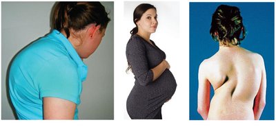
Clinical Note: Spinal Curvature Disorders
Abnormal curvatures of the vertebral column can lead to postural and functional problems.
Kyphosis: Exaggerated thoracic curvature ("hunchback").
Lordosis: Exaggerated lumbar curvature ("swayback").
Scoliosis: Lateral curvature of the spine ("crookedness").