 Back
BackThe Axial Skeleton: Structure, Function, and Key Features
Study Guide - Smart Notes
 Tailored notes based on your materials, expanded with key definitions, examples, and context.
The Axial Skeleton
Introduction to the Axial Skeleton
The axial skeleton forms the central axis of the human body and is essential for protecting vital organs and providing structural support. It includes the bones of the skull, vertebral column, and thoracic cage. The axial skeleton serves as an attachment site for muscles that move the head, neck, and back, as well as those that act across the shoulder and hip joints.
Major divisions: Skull, vertebral column, thoracic cage
Functions: Protection of the brain, spinal cord, heart, and lungs; muscle attachment; support for the body
The Skull
Major Divisions of the Skull
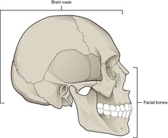
The skull is subdivided into the brain case (cranial bones) and facial bones. The brain case surrounds and protects the brain, while the facial bones support the structures of the face, form the nasal cavity, and house the teeth.
Total bones: 22 (21 immobile, 1 moveable mandible)
Brain case: 8 bones (frontal, parietal [2], temporal [2], occipital, sphenoid, ethmoid)
Facial bones: 14 bones (including maxillae, zygomatic, nasal, lacrimal, vomer, palatine, mandible)
Bones of the Brain Case
The brain case consists of flat bones that protect the brain and house structures such as the middle and inner ear. The base of the skull contains numerous openings for nerves and blood vessels.
Frontal bone: Forms the forehead and roof of the orbit.
Parietal bones: Form the upper lateral sides of the skull.
Occipital bone: Forms the posterior skull and contains the foramen magnum for the spinal cord.
Temporal bones: Form the lower lateral sides of the skull; include the mastoid and styloid processes.
Sphenoid bone: Keystone bone of the skull, forms part of the base and sides, contains the sella turcica for the pituitary gland.
Ethmoid bone: Forms the roof and lateral walls of the nasal cavity and part of the orbit.
Facial Bones
The facial bones provide the bony support for the eyes and structures of the face, including the upper and lower jaws, nose, and orbit.
Maxilla: Forms the upper jaw, hard palate, and part of the orbit; contains the infraorbital foramen.
Zygomatic bone: Cheekbone, forms part of the orbit and zygomatic arch.
Nasal bone: Forms the bridge of the nose.
Lacrimal bone: Forms part of the medial wall of the orbit.
Vomer: Forms the inferior part of the nasal septum.
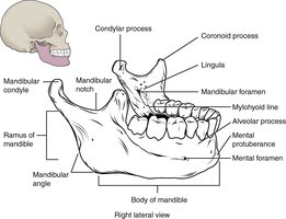
Mandible: Lower jaw, only moveable bone of the skull; contains the mental foramen and mandibular condyle.

Sutures of the Skull
Sutures are immobile fibrous joints between the bones of the skull. They are filled with dense connective tissue and provide strength and protection for the brain.
Coronal suture: Joins frontal and parietal bones.
Sagittal suture: Joins right and left parietal bones.
Lambdoid suture: Joins occipital bone to parietal and temporal bones.
Squamous suture: Joins temporal and parietal bones.
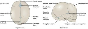
Fetal Skull and Fontanelles
The fetal skull contains large areas of dense connective tissue called fontanelles. These "soft spots" allow for flexibility during birth and growth of the brain after birth. The largest is the anterior fontanelle. Fontanelles close by age 2, but sutures remain for continued skull growth.
Anterior fontanelle: Junction of frontal and parietal bones.
Posterior, mastoid, sphenoid fontanelles: Other soft spots that close earlier.
The Vertebral Column
Regions of the Vertebral Column
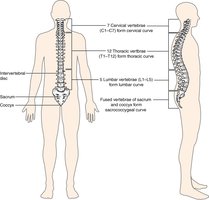
The vertebral column is divided into five regions, each with distinct vertebrae. It supports the head, protects the spinal cord, and provides attachment for ribs and muscles.
Cervical vertebrae (C1–C7): Neck region; C1 (atlas) and C2 (axis) are specialized for head movement.
Thoracic vertebrae (T1–T12): Upper back; articulate with ribs.
Lumbar vertebrae (L1–L5): Lower back; largest and bear most weight.
Sacrum: Five fused vertebrae; forms part of the pelvis.
Coccyx: Four fused vertebrae; tailbone.
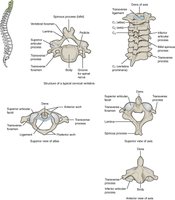
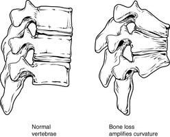
General Structure of a Vertebra
A typical vertebra consists of a body, vertebral arch (pedicles and laminae), and seven processes (spinous, transverse, superior and inferior articular). The vertebral foramen houses the spinal cord, and intervertebral discs separate adjacent vertebrae.
Body: Weight-bearing portion.
Vertebral arch: Protects the spinal cord.
Processes: Serve as muscle and ligament attachment sites.
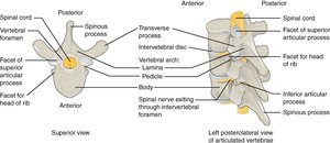
Cervical Vertebrae
Cervical vertebrae are characterized by small bodies, bifid spinous processes, and transverse foramina. The atlas (C1) supports the skull, and the axis (C2) allows for head rotation.
Atlas (C1): Ring-shaped, no body or spinous process.
Axis (C2): Has a dens (odontoid process) for rotation.
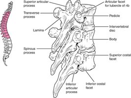
Thoracic Vertebrae
Thoracic vertebrae have larger bodies, long downward-pointing spinous processes, and facets for rib articulation. They allow for limited movement and provide attachment for the ribs.
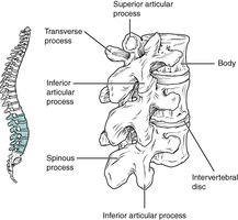
Lumbar Vertebrae
Lumbar vertebrae are the largest and strongest, with thick bodies and short, blunt spinous processes. They support the greatest amount of body weight and allow for flexion and extension of the lower back.
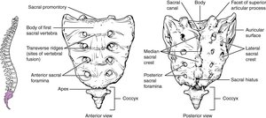
Sacrum and Coccyx
The sacrum is a triangular bone formed by the fusion of five sacral vertebrae, while the coccyx (tailbone) is formed by the fusion of four small coccygeal vertebrae. The sacrum articulates with the pelvis and supports the weight of the upper body.
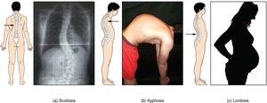
Abnormal Curvatures of the Vertebral Column
Abnormal curvatures can result from developmental anomalies, disease, or biomechanical stress. The main types are:
Kyphosis: Excessive posterior curvature of the thoracic region (humpback).
Lordosis: Excessive anterior curvature of the lumbar region (swayback).
Scoliosis: Lateral curvature with twisting of the vertebral column.
The Thoracic Cage
Structure and Function of the Thoracic Cage
The thoracic cage protects the heart and lungs and provides attachment for muscles involved in respiration and upper limb movement. It consists of the sternum, ribs, and costal cartilages.
Sternum: Manubrium, body, xiphoid process
Ribs: 12 pairs, classified as true (1–7), false (8–12), and floating (11–12)
Costal cartilages: Connect ribs to sternum
Sternum
The sternum is the elongated bone at the center of the chest, anchoring the anterior thoracic cage. It consists of three parts:
Manubrium: Superior portion, articulates with clavicles and first ribs
Body: Central portion, articulates with ribs 2–7
Xiphoid process: Inferior tip, attachment for muscles
Ribs
There are 12 pairs of ribs, each articulating posteriorly with thoracic vertebrae. The anterior attachment is via costal cartilage to the sternum (true ribs), to the cartilage of the rib above (false ribs), or not at all (floating ribs).
True ribs (1–7): Directly attached to sternum
False ribs (8–10): Indirectly attached via cartilage
Floating ribs (11–12): No anterior attachment
Summary Table: Classification of Ribs
Type | Rib Numbers | Attachment to Sternum |
|---|---|---|
True ribs | 1–7 | Direct |
False ribs | 8–10 | Indirect (via cartilage of rib above) |
Floating ribs | 11–12 | None |
Key Terms and Concepts
Foramen magnum: Large opening in the occipital bone for the spinal cord
Fontanelle: Soft spot in fetal skull for growth and flexibility
Articular process: Projection for forming joints between vertebrae
Costal cartilage: Hyaline cartilage connecting ribs to sternum
Example Application
Understanding the structure of the axial skeleton is essential for identifying bones in medical imaging, diagnosing skeletal abnormalities, and understanding the mechanics of movement and protection of vital organs.
