 Back
The Axial Skeleton: Structure, Function, and Clinical Considerations
Study Guide - Smart Notes
 Tailored notes based on your materials, expanded with key definitions, examples, and context.
The Axial Skeleton
Overview of the Skeleton
The human skeleton is a framework of bones, cartilage, joints, and ligaments that supports and protects the body. It is divided into two main divisions: the axial skeleton and the appendicular skeleton. The axial skeleton consists of 80 bones, including the skull, vertebral column, and thoracic cage, while the appendicular skeleton comprises 126 bones of the upper and lower limbs.
Axial skeleton: Skull, vertebral column, thoracic cage
Appendicular skeleton: Limbs and girdles (see Chapter 8)
The Skull
General Structure and Function
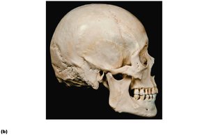
The skull is the most complex bony structure in the body, formed by cranial and facial bones. The cranial bones enclose and protect the brain and provide attachment sites for head and neck muscles. The facial bones form the framework of the face, house sense organs, provide openings for air and food, hold teeth, and anchor facial muscles.
Cranial bones: Protect the brain
Facial bones: Shape the face, support teeth, and form cavities for sense organs
Cranial Bones
The cranium is formed from eight large bones, which are classified as paired or unpaired:
Paired bones: Temporal bones, parietal bones
Unpaired bones: Frontal bone, occipital bone, sphenoid bone, ethmoid bone
Sutures of the Skull
Sutures are immovable joints that connect the bones of the skull. The four main sutures are:
Coronal suture: Between parietal and frontal bones
Squamous suture: Between parietal and temporal bones
Sagittal suture: Between right and left parietal bones
Lambdoid suture: Between parietal and occipital bones
Sutural Bones
Sutural bones are small, irregular bones found within sutures. Their presence and number vary among individuals.
Major Landmarks of the Skull
Key features of the skull include the mandibular ramus, mental foramen, alveolar processes, maxilla, zygomatic bone, nasal bone, lacrimal fossa, and others. These landmarks are important for anatomical orientation and clinical reference.
Special Parts of the Skull
Nasal Cavity and Paranasal Sinuses
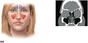
The nasal cavity is formed by several bones and is divided by the nasal septum. The paranasal sinuses are air-filled spaces within the frontal, ethmoid, sphenoid, and maxillary bones. They are lined with mucous membrane and serve to lighten the skull and enhance voice resonance.
Frontal sinus
Ethmoidal air cells
Sphenoidal sinus
Maxillary sinus
Orbits
The orbits are bony cavities that house the eyes and associated structures. They are formed by contributions from several cranial and facial bones.
The Hyoid Bone
The hyoid bone is unique in that it does not articulate directly with any other bone. It lies inferior to the mandible and serves as a movable base for the tongue, playing a crucial role in swallowing and speech.
The Vertebral Column
Structure and Function
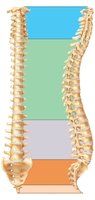
The vertebral column, or spine, is composed of 26 bones in adults. It transmits the weight of the trunk to the lower limbs, surrounds and protects the spinal cord, and provides attachment sites for muscles of the neck and back.
Cervical vertebrae (7): Neck region
Thoracic vertebrae (12): Upper back, articulate with ribs
Lumbar vertebrae (5): Lower back
Sacrum (5 fused): Articulates with hip bones
Coccyx (4 fused): Tailbone
The Thoracic Cage
Structure and Function
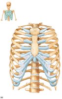
The thoracic cage forms the bony framework of the chest. It consists of the thoracic vertebrae (posteriorly), ribs (laterally), and sternum with costal cartilage (anteriorly). The thoracic cage protects thoracic organs, supports the shoulder girdle and upper limbs, and provides attachment sites for muscles.
Sternum: Manubrium, body, xiphoid process
Ribs: 12 pairs (true ribs 1–7, false ribs 8–12, floating ribs 11–12)
Sternum
The sternum is a flat bone formed from three sections: the manubrium, body, and xiphoid process. Key anatomical landmarks include the jugular notch, sternal angle, and xiphisternal joint.
Manubrium: Articulates with clavicles
Body: Articulates with costal cartilages of ribs 2–7
Xiphoid process: Ossifies around age 40
Ribs
All ribs attach posteriorly to the vertebral column. The first seven pairs are true ribs, attaching directly to the sternum via costal cartilage. The next five pairs are false ribs, with the last two pairs (11 and 12) being floating ribs that do not attach to the sternum.
Disorders of the Axial Skeleton
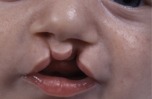
Cleft Palate
Cleft palate is a common congenital disorder in which the right and left halves of the palate fail to fuse medially, resulting in an opening in the roof of the mouth.
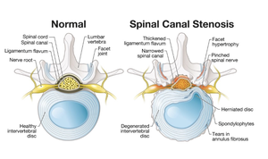
Stenosis of the Lumbar Spine
Stenosis refers to the narrowing of the vertebral canal, which can compress the roots of spinal nerves and cause pain or neurological symptoms.
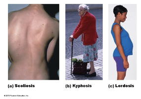
Abnormal Spinal Curvatures
Abnormal curvatures of the spine include:
Scoliosis: Abnormal lateral curvature
Kyphosis: Exaggerated thoracic curvature
Lordosis: Accentuated lumbar curvature ("swayback")
Additional info: The axial skeleton is essential for protecting the central nervous system and vital organs, supporting the body, and enabling movement. Disorders of the axial skeleton can significantly impact health and quality of life, making understanding its anatomy and function crucial for clinical practice.
