 Back
BackThe Axial Skeleton: Structure, Function, and Key Bones
Study Guide - Smart Notes
 Tailored notes based on your materials, expanded with key definitions, examples, and context.
The Skeletal System: Overview
Introduction to the Skeleton
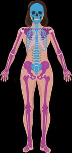
The skeletal system is composed of bones, cartilages, joints, and ligaments, providing the framework for the body and protecting vital organs. The human skeleton is divided into two main parts: the axial skeleton and the appendicular skeleton.
Axial skeleton: Includes the skull, vertebral column, and thoracic cage (80 bones). It forms the central axis of the body and protects internal organs.
Appendicular skeleton: Comprises the limbs and the pectoral and pelvic girdles (126 bones), facilitating movement and interaction with the environment.
The Skull
Introduction to the Skull
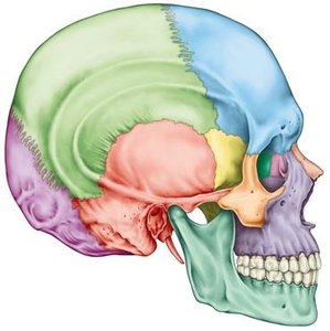
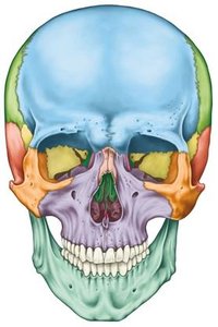
The skull is a complex bony structure that houses and protects the brain, forms the structure of the face, and provides attachment points for muscles involved in chewing and facial expression. It consists of cranial bones, facial bones, and associated bones.
Cranial bones: Form the protective case around the brain.
Facial bones: Shape the face and provide cavities for the sense organs.
Associated bones: Involved in hearing and swallowing (e.g., auditory ossicles, hyoid bone).
Cavities & Sinuses: Air-filled spaces that lighten the skull and enhance resonance of the voice.
Functions of the Skull
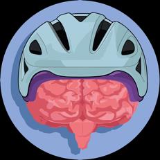
Protects the brain
Forms facial structures
Provides openings for air and food passage
Anchors teeth
Houses sensory organs (sight, hearing, taste, smell)
Major Cranial Bones
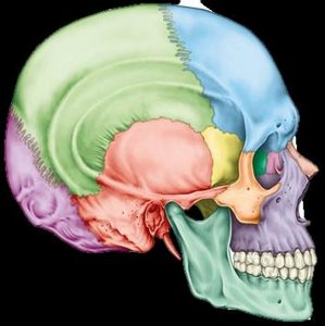
Frontal bone (1): Forehead region
Parietal bones (2): Upper sides and roof of the cranium
Occipital bone (1): Back and base of the skull; contains the foramen magnum for the spinal cord
Temporal bones (2): Sides of the skull, house structures for hearing (auditory meatus, styloid, mastoid, and zygomatic processes)
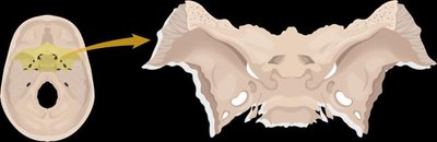
Sphenoid bone (1): Butterfly-shaped bone at the base of the skull; contains the sella turcica, which houses the pituitary gland
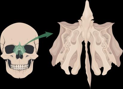
Ethmoid bone (1): Light, spongy bone between the orbits and nasal cavity; includes the crista galli and cribriform plate for olfactory nerves
Major Facial Bones
Maxilla (2): Upper jaw, forms part of the orbit, nasal cavity, and hard palate
Zygomatic (2): Cheekbones
Nasal (2): Bridge of the nose
Lacrimal (2): Medial wall of the orbit
Inferior Nasal Conchae (2): Lateral walls of the nasal cavity
Vomer (1): Inferior part of the nasal septum
Palatine (2): Posterior part of the hard palate
Mandible (1): Lower jaw; only freely movable bone of the skull
Cavities and Sinuses of the Skull
The skull contains several cavities and sinuses that serve important functions:
Orbital cavity: Houses the eyes; formed by the frontal, sphenoid, zygomatic, maxilla, lacrimal, ethmoid, and palatine bones.
Nasal cavity: Formed by the maxilla, nasal, sphenoid, vomer, inferior nasal conchae, palatine, and ethmoid bones.
Paranasal sinuses: Air-filled spaces in the frontal, ethmoid, maxilla, and sphenoid bones; lighten the skull, warm and moisten air, and enhance vocal resonance.
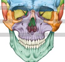
Table: Paranasal Sinuses and Their Locations
Sinus | Location |
|---|---|
Frontal sinus | Frontal bone (above the eyes) |
Ethmoid air cells | Ethmoid bone (between the eyes) |
Maxillary sinus | Maxilla (cheek area) |
Sphenoidal sinus | Sphenoid bone (deep to the nose) |
The Vertebral Column (Spine)
Structure and Regions of the Spine
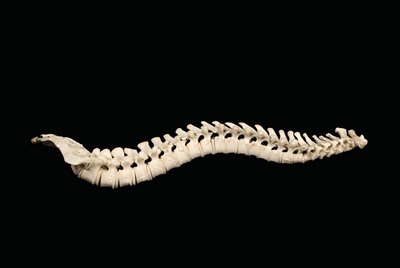
The vertebral column supports the body, protects the spinal cord, and provides attachment points for ribs and muscles. It consists of 24 vertebrae, the sacrum, and the coccyx, divided into five regions:
Cervical (7): Neck region (C1–C7); C1 (atlas) supports the head, C2 (axis) allows rotation.
Thoracic (12): Upper back (T1–T12); articulate with ribs.
Lumbar (5): Lower back (L1–L5); largest vertebrae for weight-bearing.
Sacrum (5 fused): Forms the posterior wall of the pelvis.
Coccyx (3–5 fused): Tailbone.
Spinal Curvatures and Intervertebral Discs
Four curvatures: cervical, thoracic, lumbar, and sacral; help absorb shock and maintain balance.
Intervertebral discs: fibrocartilaginous pads between vertebrae, providing cushioning and flexibility (absent between sacrum and coccyx).
The Thoracic Cage
Structure and Function
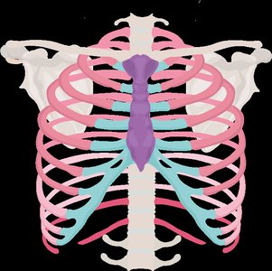
The thoracic cage protects the heart and lungs and supports the shoulder girdles and upper limbs. It consists of the thoracic vertebrae, ribs, and sternum.
Ribs (12 pairs): Flat bones that wrap around the chest.
True ribs (1–7): Attach directly to the sternum via costal cartilage.
False ribs (8–12): Attach indirectly or not at all to the sternum; ribs 11–12 are floating ribs with no sternal attachment.
Sternum: Flat bone consisting of the manubrium, body, and xiphoid process.
Intercostal cartilage: Connects ribs 1–10 to the sternum, allowing flexibility during breathing.
The Pectoral Girdle
Structure and Function
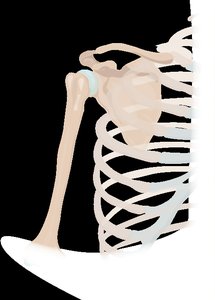
The pectoral (shoulder) girdle attaches the upper limbs to the axial skeleton and allows for a wide range of motion. It consists of the clavicle and scapula.
Clavicle: Collarbone; articulates with the sternum and scapula.
Scapula: Shoulder blade; articulates with the clavicle and humerus. Key features include the acromion process (articulates with clavicle) and glenoid cavity (shoulder socket for the humerus).
Bones of the Upper Limb
Arm, Forearm, Wrist, and Hand
Humerus: Single bone of the upper arm; articulates with the scapula at the shoulder and with the radius and ulna at the elbow.
Radius: Lateral bone of the forearm (thumb side); allows rotation of the forearm.
Ulna: Medial bone of the forearm (pinky side); forms the elbow joint with the humerus.
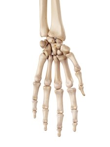
Carpals (8): Short bones of the wrist.
Metacarpals (5): Long bones of the palm, numbered I (thumb) to V (pinky).
Phalanges (14): Bones of the fingers; each finger has three (proximal, middle, distal), except the thumb, which has two.
The Pelvic Girdle
Structure and Function
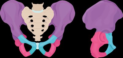
The pelvic girdle attaches the lower limbs to the axial skeleton and supports the weight of the upper body. It consists of two coxal (hip) bones, each formed by the fusion of three bones: ilium, ischium, and pubis.
Ilium: Superior, flared region; forms the iliac crest.
Ischium: Inferior, posterior region; supports body weight when sitting.
Pubis: Inferior, anterior region; joined at the pubic symphysis.
Acetabulum: Deep socket formed by all three bones; articulates with the femur.
Differences Between Male and Female Pelvis
Feature | Male | Female |
|---|---|---|
General Appearance | Narrow & robust | Broader & lighter |
Angle of Pubic Arch | More acute | Wider, rounder |
Shape of Pelvic Inlet | Heart-shaped, closer together | Oval, further apart |
Note: The female pelvis is adapted for childbirth.
Bones of the Lower Limb
Thigh, Leg, Ankle, and Foot
Femur: Longest and strongest bone; articulates with the acetabulum, tibia, and patella.
Patella: Kneecap; a sesamoid bone within the quadriceps tendon.
Tibia: Larger, medial bone of the lower leg; bears most of the body’s weight.
Fibula: Slender, lateral bone; stabilizes the ankle.
Tarsals (7): Short bones of the ankle; includes the talus (articulates with tibia) and calcaneus (heel bone).
Metatarsals (5): Long bones of the foot, forming the arches.
Phalanges (14): Bones of the toes; each toe has three, except the big toe, which has two.
Summary Table: Major Divisions of the Skeleton
Division | Main Components | Number of Bones | Primary Function |
|---|---|---|---|
Axial Skeleton | Skull, vertebral column, thoracic cage | 80 | Support, protection of organs |
Appendicular Skeleton | Limbs, pectoral & pelvic girdles | 126 | Movement, manipulation of environment |
