 Back
BackThe Axial Skeleton: Structure, Function, and Key Features
Study Guide - Smart Notes
 Tailored notes based on your materials, expanded with key definitions, examples, and context.
The Axial Skeleton
Overview and Divisions
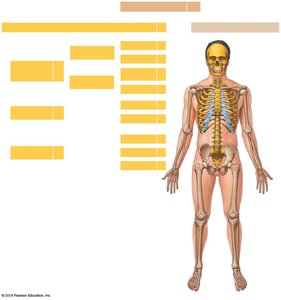
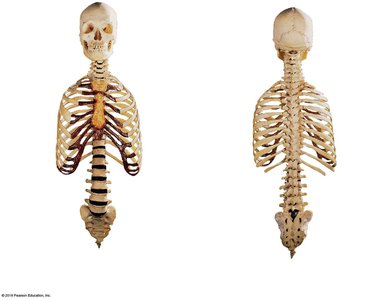
The axial skeleton forms the central axis of the human body and consists of 80 bones. It includes the skull, vertebral column, and thoracic cage. The axial skeleton supports and protects the organs in the dorsal and ventral body cavities and provides attachment points for muscles involved in head, neck, and trunk movements, as well as respiration.
Axial Skeleton: Bones of the head and trunk (skull, vertebral column, thoracic cage).
Appendicular Skeleton: Bones that support the limbs (not covered in this chapter).
/
Functions of the Axial Skeleton:
Supports and protects organs in body cavities
Provides attachment points for muscles that adjust the head, neck, and trunk, perform breathing movements, and stabilize the appendicular skeleton
The Skull
Structure and Subdivisions
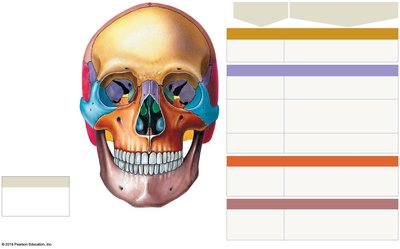
The skull is composed of 22 bones: 8 cranial bones and 14 facial bones. The cranial bones form the cranium, which protects the brain, while the facial bones support the entrances to the digestive and respiratory tracts.
Cranial Bones (8): Frontal, parietal (2), temporal (2), occipital, sphenoid, ethmoid
Facial Bones (14): Maxillae (2), palatine (2), nasal (2), inferior nasal conchae (2), zygomatic (2), lacrimal (2), vomer, mandible
Associated Bones: Hyoid bone, auditory ossicles (6)
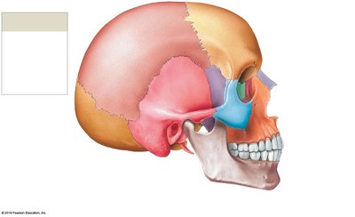
Sutures of the Skull
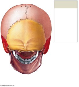
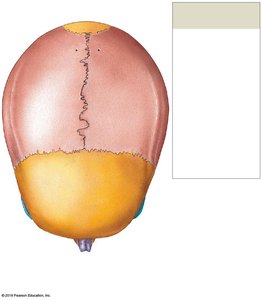
Sutures are immovable joints that connect the bones of the skull. The four major sutures are:
Lambdoid Suture: Separates occipital from parietal bones
Coronal Suture: Attaches frontal bone to parietal bones
Sagittal Suture: Between parietal bones, from lambdoid to coronal suture
Squamous Sutures: Join temporal bones with parietal bones
Sinuses
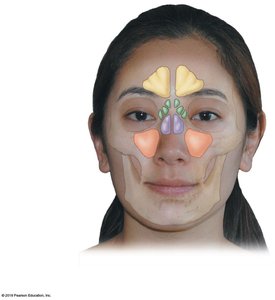
Sinuses are air-filled chambers within the skull that decrease its weight, are lined with mucous membranes, and serve as resonating chambers for speech.
Cranial Bones: Structure and Features
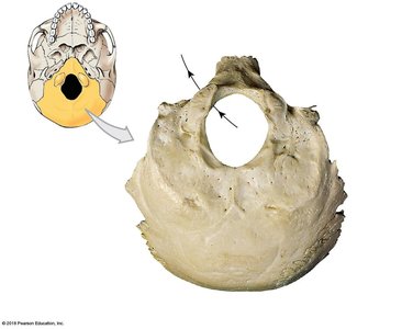
Occipital Bone
Function: Forms much of the posterior and inferior surfaces of the cranium
Key Markings: Occipital condyles (articulate with the atlas), foramen magnum (connects cranial cavity with vertebral canal), external occipital protuberance
Parietal Bones
Function: Form part of the superior and lateral surfaces of the cranium
Key Markings: Superior and inferior temporal lines (muscle attachment), grooves for cranial blood vessels
' OW notar woah! 1 evil \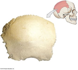
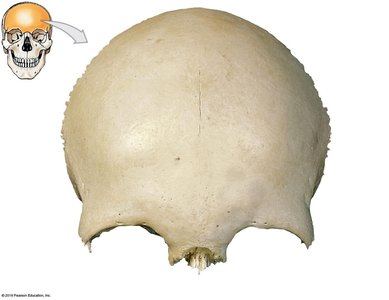
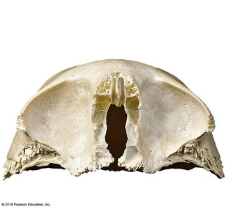
Frontal Bone
Function: Forms the anterior cranium and roof of the orbits; contains frontal sinuses
Key Markings: Forehead, supra-orbital margin, glabella, lacrimal fossa
Foramina: Supra-orbital foramen (for blood vessels and nerves)
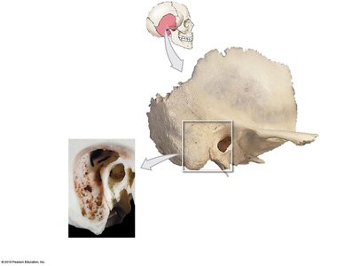
Temporal Bones
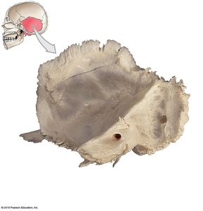
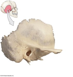
Function: Form parts of the lateral cranium and zygomatic arches, articulate with the mandible, surround and protect the internal ear
Key Markings: Squamous part, zygomatic process, mandibular fossa, mastoid process, styloid process, petrous part
Foramina: Carotid canal, external acoustic meatus, stylomastoid foramen, internal acoustic meatus
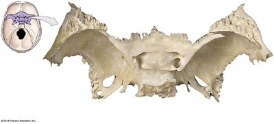
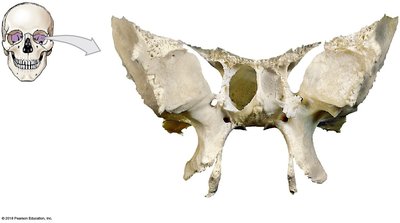
Sphenoid
Function: Forms part of the floor of the cranium, unites cranial and facial bones, contains sphenoidal sinuses
Key Markings: Body, sella turcica, hypophyseal fossa, lesser and greater wings, pterygoid processes
Foramina: Optic canals, superior orbital fissure, foramen rotundum, foramen ovale, foramen spinosum
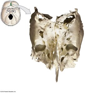
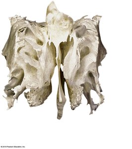
Ethmoid
Function: Forms the anteromedial floor of the cranium, roof of the nasal cavity, part of the nasal septum, and medial orbital wall
Key Markings: Cribriform plate, crista galli, ethmoidal labyrinths, perpendicular plate
Foramina: Olfactory foramina (for olfactory nerves)
Facial Bones: Structure and Features
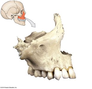
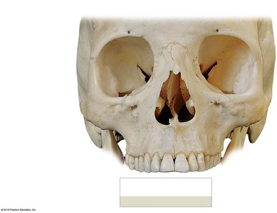
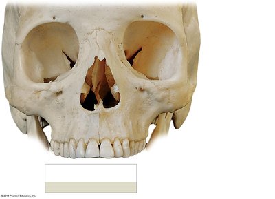
Maxillae
Function: Support upper teeth, form the upper jaw, hard palate, and parts of the orbit and nasal cavity
Key Markings: Orbital rim, anterior nasal spine, alveolar process, palatine processes, maxillary sinuses
Foramina: Infra-orbital foramen, inferior orbital fissure
Palatine Bones
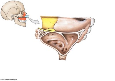
Function: Form posterior portions of the hard palate and contribute to the floors of the orbits
Key Markings: Horizontal plate, perpendicular plate
Nasal Bones, Vomer, Inferior Nasal Conchae, Zygomatic Bones, Lacrimal Bones
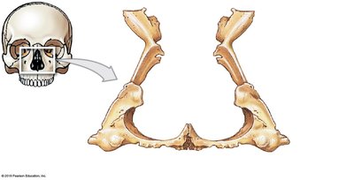
Nasal Bones: Support the bridge of the nose
Vomer: Forms the inferior portion of the bony nasal septum
Inferior Nasal Conchae: Create air turbulence and increase surface area in the nasal cavity
Zygomatic Bones: Form the lateral wall of the orbit and part of the zygomatic arch
Lacrimal Bones: Form part of the medial wall of the orbit; contain the lacrimal sulcus
Mandible
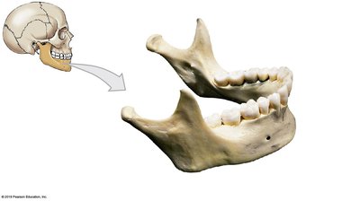
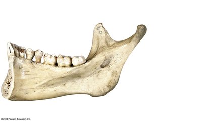
Function: Forms the lower jaw
Key Markings: Body, alveolar part, mental protuberance, ramus, condylar process, coronoid process, mandibular notch
Foramina: Mental foramina, mandibular foramen
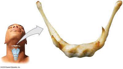
Hyoid Bone
Function: Supports the larynx and attaches muscles of the larynx, pharynx, and tongue
Key Markings: Body, greater horns, lesser horns
Orbital and Nasal Complexes
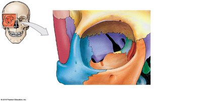
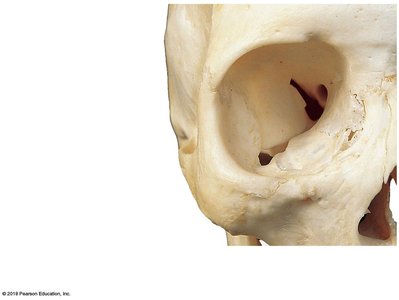
Orbital Complex
The orbits are formed by seven bones: frontal, maxilla, lacrimal, ethmoid, sphenoid, palatine, and zygomatic. These bones protect the eyes and associated structures.
Nasal Complex and Paranasal Sinuses
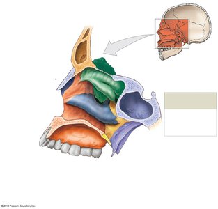
The nasal complex is formed by bones that enclose the nasal cavities and paranasal sinuses. Paranasal sinuses are air-filled chambers that lighten the skull and produce mucus.
Fontanelles and Infant Skull
Fontanelles
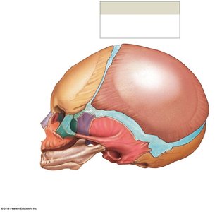
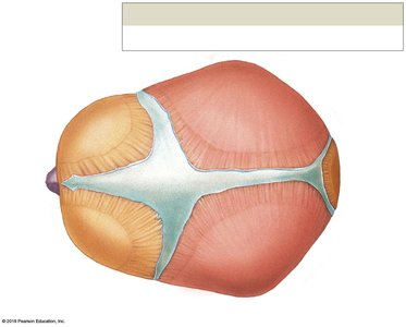
Fontanelles are large areas of fibrous connective tissue in the infant skull that cover unfused sutures. They allow the skull to flex during birth and accommodate rapid brain growth.
Sphenoidal Fontanelles: At the junction of squamous and coronal sutures
Mastoid Fontanelles: At the junction of squamous and lambdoid sutures
Anterior Fontanelle: At the intersection of frontal, sagittal, and coronal sutures
Posterior Fontanelle: At the junction of lambdoid and sagittal sutures
The Vertebral Column
Structure and Curves
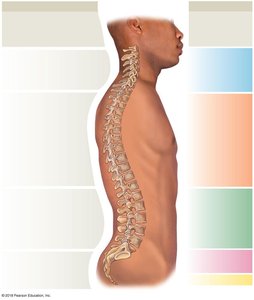
The vertebral column (spine) protects the spinal cord and supports the head and body. It consists of 26 bones: 24 vertebrae, the sacrum, and the coccyx. The column has four major curves: cervical, thoracic, lumbar, and sacral.
Primary Curves: Thoracic and sacral (present during fetal development)
Secondary Curves: Cervical and lumbar (develop after birth)
Vertebral Regions
Cervical (C1–C7): Neck region; small bodies, large vertebral foramen
Thoracic (T1–T12): Upper back; articulate with ribs; heart-shaped bodies
Lumbar (L1–L5): Lower back; largest vertebrae; thick, oval-shaped bodies
Sacral (Sacrum): Five fused vertebrae; articulates with pelvic girdle
Coccygeal (Coccyx): Three to five fused vertebrae; tailbone
General Vertebral Anatomy
Vertebral Body: Transfers weight along the spine
Vertebral Arch: Forms the posterior margin of the vertebral foramen
Articular Processes: Lateral projections for articulation with adjacent vertebrae
Spinous and Transverse Processes: Muscle and ligament attachment
Intervertebral Discs: Pads of fibrocartilage separating vertebral bodies
Intervertebral Foramina: Gaps for nerve connections to the spinal cord
The Thoracic Cage
Structure and Function
The thoracic cage consists of the thoracic vertebrae, ribs, costal cartilages, and sternum. It protects the heart, lungs, and thymus, and provides attachment for muscles involved in breathing and movement of the pectoral girdles.
Ribs: 12 pairs; true ribs (1–7) attach directly to the sternum, false ribs (8–12) do not
Floating Ribs: Ribs 11–12; do not attach to the sternum
Sternum: Flat bone with three parts—manubrium, body, and xiphoid process
Key Functions:
Protects thoracic organs
Supports breathing movements
Provides attachment for muscles of the back, chest, and shoulders
Summary Table: Major Bones of the Axial Skeleton
Region | Bones | Key Functions |
|---|---|---|
Skull | 22 (8 cranial, 14 facial) | Protects brain, supports sensory structures |
Auditory Ossicles | 6 | Transmit sound vibrations |
Hyoid | 1 | Supports larynx, tongue movement |
Vertebral Column | 26 (24 vertebrae, sacrum, coccyx) | Protects spinal cord, supports body |
Thoracic Cage | 25 (24 ribs, sternum) | Protects thoracic organs, aids respiration |
Additional info: The axial skeleton is essential for protecting the central nervous system and vital organs, and for providing structural support and leverage for movement. Understanding its anatomy is foundational for further study in anatomy, physiology, and clinical practice.
