 Back
BackThe Brain and Cranial Nerves: Structure, Development, and Function
Study Guide - Smart Notes
 Tailored notes based on your materials, expanded with key definitions, examples, and context.
The Brain and Cranial Nerves
Introduction to Brain Structure and Function
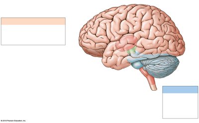
The adult human brain is a complex organ responsible for integrating sensory information, coordinating motor output, and supporting higher mental functions such as thought, memory, and emotion. It is divided into several major regions, each with specialized roles.
Cerebrum: Controls conscious thought, intellect, memory, and voluntary muscle activity.
Cerebellum: Coordinates complex somatic motor patterns and maintains posture and balance.
Diencephalon: Includes the thalamus (sensory relay), hypothalamus (autonomic and endocrine control), and pituitary gland.
Brainstem: Composed of the midbrain, pons, and medulla oblongata; relays information and regulates vital functions.
Major Brain Regions, Vesicles, and Ventricles
Embryonic Development of the Brain
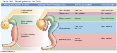
The brain develops from the neural tube, forming three primary brain vesicles which further differentiate into secondary vesicles and mature brain regions. This organization is crucial for understanding the adult brain's structure and ventricular system.
Primary Vesicle (3 weeks) | Secondary Vesicle (6 weeks) | Brain Region at Birth | Ventricular System |
|---|---|---|---|
Prosencephalon (Forebrain) | Telencephalon | Cerebrum | Lateral ventricles |
Diencephalon | Diencephalon | Third ventricle | |
Mesencephalon (Midbrain) | Mesencephalon | Midbrain | Cerebral aqueduct |
Rhombencephalon (Hindbrain) | Metencephalon | Cerebellum and Pons | Fourth ventricle |
Myelencephalon | Medulla oblongata | Fourth ventricle |
Ventricular System of the Brain
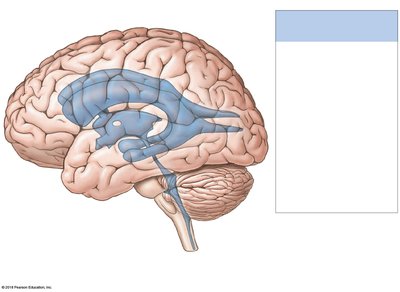
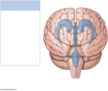
The brain contains interconnected chambers called ventricles, filled with cerebrospinal fluid (CSF). These include the paired lateral ventricles, third ventricle, and fourth ventricle, which are essential for cushioning the brain and circulating nutrients.
Lateral ventricles: Located in each cerebral hemisphere.
Third ventricle: Located in the diencephalon, connected to lateral ventricles via interventricular foramina.
Fourth ventricle: Located between the pons and cerebellum, continuous with the central canal of the spinal cord.
Protection and Support of the Brain
Cranial Meninges and Dural Folds
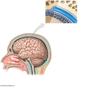
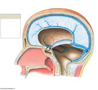
The brain is protected by the bones of the cranium, three layers of cranial meninges (dura mater, arachnoid mater, pia mater), and cerebrospinal fluid. Dural folds (falx cerebri, tentorium cerebelli, falx cerebelli) stabilize and support the brain within the cranial cavity.
Dura mater: Tough outer layer with periosteal and meningeal components.
Arachnoid mater: Middle layer, contains the subarachnoid space filled with CSF.
Pia mater: Thin inner layer, adheres to the brain surface.
Cerebrospinal Fluid (CSF)
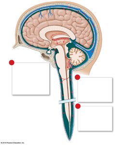
CSF surrounds the CNS, providing mechanical protection, supporting the brain, and transporting nutrients and waste. It is produced by the choroid plexus and circulates through the ventricles, central canal, and subarachnoid space, eventually being absorbed into venous circulation via arachnoid granulations.
Functions: Cushioning, nutrient transport, waste removal.
Production: Choroid plexus (specialized ependymal cells and capillaries).
Circulation: Lateral ventricles → third ventricle → fourth ventricle → subarachnoid space → venous system.
Brainstem: Medulla Oblongata, Pons, and Midbrain
Medulla Oblongata
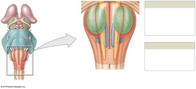
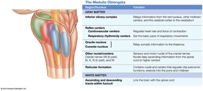
The medulla oblongata is the most inferior part of the brainstem, connecting the brain to the spinal cord. It contains nuclei that regulate autonomic functions (cardiovascular and respiratory centers), relay sensory and motor information, and house cranial nerve nuclei.
Autonomic centers: Control heart rate, blood pressure, and digestion.
Relay stations: Gracile and cuneate nuclei relay sensory information.
Cranial nerves: VIII, IX, X, XI, XII originate here.
Region/Nucleus | Function |
|---|---|
Inferior olivary complex | Relays information to the cerebellum |
Cardiovascular centers | Regulate heart rate and force of contraction |
Respiratory rhythmicity centers | Set the basic pace of respiratory movements |
Gracile and cuneate nuclei | Relay somatic sensory information to the thalamus |
Reticular formation | Contains nuclei and centers that regulate vital autonomic functions |
Ascending and descending tracts | Link the brain with the spinal cord |
Pons
The pons is located superior to the medulla and contains nuclei involved in respiration, sensory and motor relay to the cerebellum, and cranial nerve nuclei. It acts as a bridge between different parts of the nervous system.
Respiratory centers: Apneustic and pneumotaxic centers regulate breathing.
Tracts: Ascending, descending, and transverse fibers connect the pons with other brain regions.
Cranial nerves: V, VI, VII, VIII originate here.
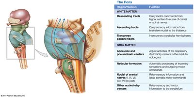
Region/Nucleus | Function |
|---|---|
Descending tracts | Carry motor commands from higher centers |
Ascending tracts | Carry sensory information to the thalamus |
Transverse pontine fibers | Interconnect cerebellar hemispheres |
Apneustic and pneumotaxic centers | Adjust respiratory rhythmicity |
Reticular formation | Automatic processing of incoming sensations and outgoing motor commands |
Midbrain
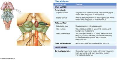
The midbrain (mesencephalon) is involved in processing visual and auditory information, maintaining consciousness, and controlling reflexive motor responses. It contains the tectum (corpora quadrigemina), tegmentum (red nucleus, substantia nigra), and cerebral peduncles.
Superior colliculi: Visual reflexes.
Inferior colliculi: Auditory reflexes.
Red nucleus and substantia nigra: Motor coordination and regulation.
Region/Nucleus | Function |
|---|---|
Superior colliculi | Integrate visual information and initiate reflexes |
Inferior colliculi | Relay auditory information and initiate reflexes |
Red nucleus | Regulates upper limb position and background muscle tone |
Substantia nigra | Regulates activity in the basal nuclei |
Cerebral peduncles | Connect primary motor cortex with motor neurons in brain and spinal cord |
The Cerebellum
Structure and Function
The cerebellum is responsible for coordinating voluntary movements, maintaining posture and balance, and fine-tuning motor activity. It consists of two hemispheres, a central vermis, and three lobes (anterior, posterior, flocculonodular).
Cerebellar cortex: Gray matter with folia (folds) for increased surface area.
Arbor vitae: Internal white matter structure.
Purkinje cells: Large neurons that integrate sensory and motor information.
Cerebellar peduncles: Superior, middle, and inferior tracts connecting the cerebellum to the brainstem.
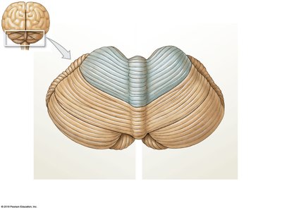
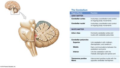
Region/Nucleus | Function |
|---|---|
Cerebellar cortex | Coordination and control of ongoing body movements |
Cerebellar nuclei | Involuntary coordination and control |
Arbor vitae | Connects cortex and nuclei with cerebellar peduncles |
Peduncles (superior, middle, inferior) | Link cerebellum with other brain regions and spinal cord |
The Diencephalon
Thalamus
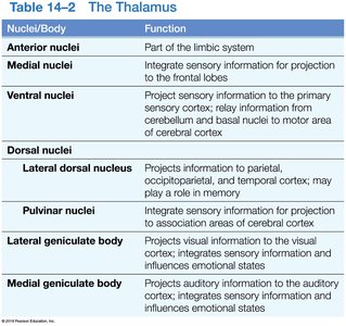
The thalamus acts as a relay and processing center for sensory information, directing it to appropriate areas of the cerebral cortex. It consists of several nuclei with specialized functions.
Nuclei/Body | Function |
|---|---|
Anterior nuclei | Part of the limbic system |
Medial nuclei | Integrate sensory information for projection to the frontal lobes |
Ventral nuclei | Project sensory information to the primary sensory cortex |
Lateral dorsal nucleus | Projects to parietal, occipitoparietal, and temporal cortex |
Pulvinar nuclei | Integrate sensory information for projection to association areas |
Lateral geniculate body | Visual information relay |
Medial geniculate body | Auditory information relay |
Hypothalamus
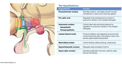
The hypothalamus is a key regulator of homeostasis, controlling autonomic functions, hormone production, emotional responses, and circadian rhythms. It connects to the pituitary gland via the infundibulum.
Region/Nucleus | Function |
|---|---|
Paraventricular nucleus | Secretes oxytocin, stimulates smooth muscle contractions |
Pre-optic area | Regulates body temperature |
Autonomic centers | Control heart rate and blood pressure |
Lateral tuberal nuclei | Produce hormones that control pituitary gland secretion |
Mammillary bodies | Control feeding reflexes |
Supra-optic nucleus | Secretes antidiuretic hormone (ADH) |
Suprachiasmatic nucleus | Regulates circadian rhythms |
The Limbic System
Structure and Function
The limbic system is a functional grouping of structures involved in emotion, motivation, and memory. It links conscious and unconscious brain functions and facilitates memory storage and retrieval.
Limbic lobe: Cingulate gyrus, parahippocampal gyrus, dentate gyrus, hippocampus.
Amygdaloid body: Interface between limbic system, cerebrum, and sensory systems.
Fornix: White matter tract connecting hippocampus to hypothalamus.
Anterior thalamic nuclei: Relay information within the limbic system.
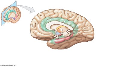
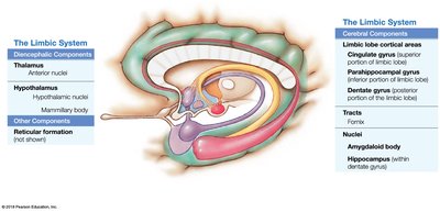
Component | Function |
|---|---|
Cingulate gyrus | Emotion formation and processing |
Hippocampus | Memory formation |
Amygdaloid body | Emotional responses, fear, aggression |
The Cerebrum
Major Anatomical Subdivisions and Functions
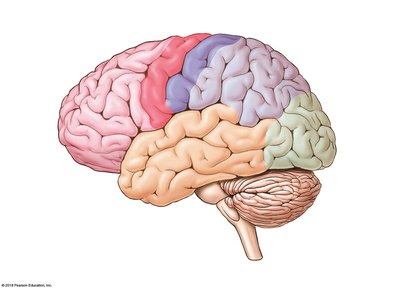
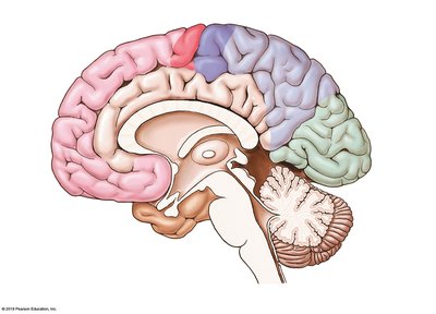
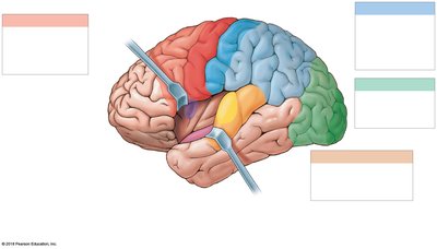
The cerebrum is divided into two hemispheres and four main lobes, each with specialized functions. It is responsible for all conscious thought, voluntary movement, and sensory processing.
Frontal lobe: Voluntary motor control, planning, judgment, speech (Broca's area).
Parietal lobe: Sensory perception and integration.
Temporal lobe: Hearing, memory, language comprehension (Wernicke's area).
Occipital lobe: Vision and visual processing.
White Matter and Basal Nuclei
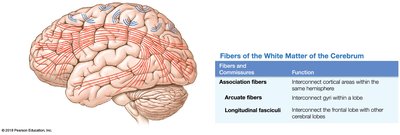
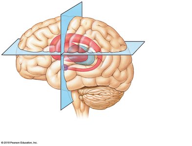
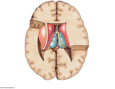
White matter consists of myelinated axons connecting different brain regions. Basal nuclei are deep gray matter structures involved in the regulation of movement and muscle tone.
Association fibers: Connect areas within the same hemisphere.
Commissural fibers: Connect the two hemispheres (e.g., corpus callosum).
Projection fibers: Connect the cerebrum with lower brain regions and spinal cord.
Basal nuclei: Caudate nucleus, putamen, globus pallidus, claustrum; regulate movement.
Functional Areas of the Cerebral Cortex
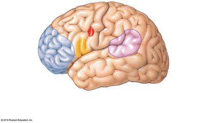
The cerebral cortex contains specialized areas for motor, sensory, and association functions. Each lobe contains primary and association areas for processing specific types of information.
Lobe/Area | Function |
|---|---|
Primary motor cortex (frontal lobe) | Voluntary control of skeletal muscles |
Primary somatosensory cortex (parietal lobe) | Conscious perception of touch, pressure, pain, vibration, temperature |
Visual cortex (occipital lobe) | Conscious perception of visual stimuli |
Auditory and olfactory cortex (temporal lobe) | Conscious perception of auditory and olfactory stimuli |
Association areas | Integration and processing of sensory and motor information |
Brain Waves and Electroencephalogram (EEG)
Brain activity can be measured using an EEG, which records electrical patterns known as brain waves. Different types of brain waves are associated with various states of consciousness and brain function.
Alpha waves: Relaxed, awake adults with eyes closed.
Beta waves: Active, alert, or stressed adults.
Theta waves: Children, frustrated adults, or brain disorders.
Delta waves: Deep sleep, infants, or brain damage in adults.
Cranial Nerves and Cranial Reflexes
Cranial Nerves
There are 12 pairs of cranial nerves, each with specific sensory, motor, or mixed functions. They are essential for sensory perception, motor control, and autonomic regulation in the head and neck.
Olfactory (I): Smell
Optic (II): Vision
Oculomotor (III), Trochlear (IV), Abducens (VI): Eye movements
Trigeminal (V): Sensation and mastication
Facial (VII): Facial expression, taste
Vestibulocochlear (VIII): Hearing and balance
Glossopharyngeal (IX): Taste, swallowing
Vagus (X): Autonomic control of thoracic and abdominal organs
Accessory (XI): Head and neck movement
Hypoglossal (XII): Tongue movement
Cranial Reflexes
Cranial reflexes are rapid, involuntary responses involving cranial nerves and brainstem integration centers. They are important for clinical assessment of brainstem function.
Pupillary light reflex: Pupil constriction in response to light (CN II → CN III).
Corneal (blink) reflex: Blinking when the cornea is touched (CN V → CN VII).
Gag reflex: Gagging when the posterior pharynx is stimulated (CN IX → CN X).
Vestibulo-ocular reflex (VOR): Eye stabilization during head movement (CN VIII → CN III, IV, VI).
Lacrimation reflex: Tear production in response to eye irritation.
Jaw-jerk reflex: Jaw contraction when the chin is tapped (CN V for both afferent and efferent).
Clinical significance: Testing cranial reflexes is essential in neurological exams to assess brainstem integrity and diagnose potential lesions or brainstem death.
