 Back
BackThe Brain and Cranial Nerves: Structure and Function of the Cerebrum
Study Guide - Smart Notes
 Tailored notes based on your materials, expanded with key definitions, examples, and context.
Introduction to the Central Nervous System (CNS)
Overview of the CNS
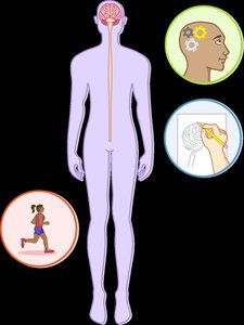
The central nervous system (CNS) is composed of two main components: the brain and the spinal cord. It serves as the primary integration and command center for the human body, responsible for processing sensory information, coordinating movement, and higher cognitive functions such as thought, memory, and emotion.
Brain: The control center for processing and interpreting sensory information and directing responses.
Spinal Cord: Conducts signals to and from the brain and controls reflex activities.
Functions: Involved in memory, voluntary movement, decision-making, and all conscious and unconscious activities.
Major Regions of the Brain
Divisions of the Adult Brain
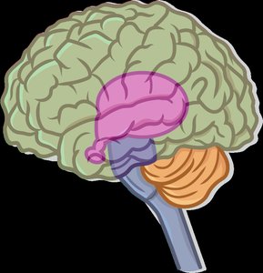
The adult brain is divided into four major regions, each with specialized functions:
Cerebrum: Largest, most anterior part; responsible for higher cognitive functions.
Diencephalon: Central part of the forebrain; includes the thalamus, hypothalamus, and epithalamus.
Brainstem: Connects the cerebrum to the spinal cord; consists of the midbrain, pons, and medulla oblongata.
Cerebellum: Located at the back of the brain; coordinates motor activity and balance.
CNS Development
Embryonic Origins of the CNS
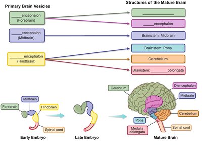
The CNS develops from the neural tube during embryogenesis. The anterior end of the neural tube forms three primary brain vesicles, which further differentiate into the major regions of the mature brain.
Primary Brain Vesicles: Prosencephalon (forebrain), mesencephalon (midbrain), rhombencephalon (hindbrain).
Mature Structures: The prosencephalon becomes the cerebrum and diencephalon; the mesencephalon forms the midbrain; the rhombencephalon forms the pons, cerebellum, and medulla oblongata.
Spinal Cord: Develops from the caudal end of the neural tube.
White and Gray Matter
Organization of Nervous Tissue
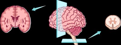
The brain and spinal cord are composed of two types of nervous tissue: white matter and gray matter.
White Matter: Consists mainly of myelinated axons; responsible for rapid signal transmission.
Gray Matter: Contains neuron cell bodies, dendrites, and unmyelinated axons; involved in processing and integration.
Brain: Outer layer of gray matter (cortex), inner white matter, and subcortical gray matter nuclei.
Spinal Cord: Outer white matter, inner gray matter.
The Cerebrum
Structure and Functional Organization
The cerebrum consists of two cerebral hemispheres and is responsible for higher-order functions such as reasoning, language, and voluntary movement. Each hemisphere is specialized for certain functions (lateralization), and controls the opposite side of the body (contralateral control).
Left Hemisphere: Language, intellect, logical thinking.
Right Hemisphere: Visual-spatial skills, emotion, artistic/musical abilities.
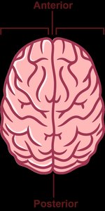
Surface Features of the Cerebrum
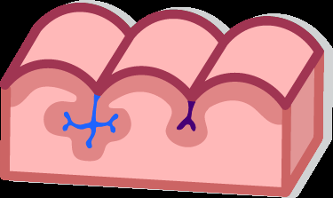
The surface of the cerebrum is marked by elevated ridges (gyri), shallow grooves (sulci), and deep grooves (fissures), which increase surface area for cortical processing.
Gyri: Elevated ridges of tissue.
Sulci: Shallow grooves separating gyri.
Fissures: Deep grooves separating larger brain regions.
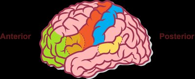
Lobes of the Cerebrum
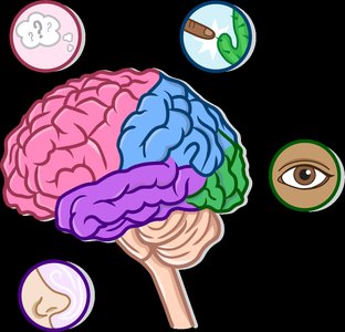
The cerebrum is divided into four lobes, each named after the cranial bone that overlies it and associated with specific functions:
Frontal Lobe: Voluntary movement, planning, decision-making, personality.
Parietal Lobe: Sensation, spatial perception, temperature, pain, touch.
Occipital Lobe: Vision and visual association.
Temporal Lobe: Hearing, smell, memory.
Functional Areas of the Cerebral Cortex
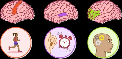
The cerebral cortex contains three main types of functional areas:
Motor Areas: Control voluntary movement.
Sensory Areas: Receive and process sensory input.
Association Areas: Integrate information for complex processes such as recognition, memory, and reasoning.
Motor and sensory areas are primarily concerned with the contralateral side of the body.
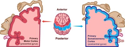
Primary Motor Cortex & Primary Somatosensory Cortex
The primary motor cortex (in the precentral gyrus) initiates voluntary movements, while the primary somatosensory cortex (in the postcentral gyrus) receives sensory information from the body. The homunculus is a visual representation showing the relative amount of cortex devoted to different body parts, reflecting the density of neural connections.
Larger area on the motor homunculus = more precise motor control.
Larger area on the sensory homunculus = greater sensitivity.
Specialized Areas of the Cerebral Cortex
Several specialized regions are critical for language, reasoning, and voluntary movement:
Wernicke’s Area: Language comprehension; located in the temporal lobe.
Broca’s Area: Speech production; located in the frontal lobe.
Prefrontal Cortex: Intellect, cognition, personality, reasoning, and planning; most anterior part of the cerebrum.

Cerebral White Matter
Types of White Matter Fibers
Cerebral white matter is responsible for communication within the brain and between the brain and spinal cord. It is classified into three groups based on the direction of fibers:
Association Fibers: Connect different cortical areas within the same hemisphere.
Commissural Fibers: Connect corresponding areas of the two hemispheres (e.g., corpus callosum).
Projection Fibers: Connect the cerebrum with lower brain regions and the spinal cord (e.g., corona radiata, internal capsule).
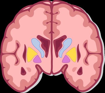
Basal Nuclei (Basal Ganglia)
Structure and Function
The basal nuclei (also called basal ganglia) are clusters of subcortical neuron cell bodies that communicate with the premotor cortex. They are primarily responsible for regulating movement, including starting and stopping motions and inhibiting unnecessary movements. Dysfunction of the basal nuclei is associated with movement disorders such as Parkinson’s disease.
Main Components: Caudate nucleus, putamen, globus pallidus (collectively called the striatum).
Function: Smooth, coordinated voluntary movement.
White Matter | Gray Matter |
|---|---|
Myelinated axons | Neuron cell bodies, dendrites, unmyelinated axons |
Signal transmission | Information processing |
Inner brain, outer spinal cord | Outer brain (cortex), inner spinal cord |
Additional info: The notes above expand on the provided content with academic context, definitions, and examples to ensure a comprehensive, self-contained study guide for ANP college students.
