 Back
BackThe Brain and Cranial Nerves: Structure, Function, and Integration
Study Guide - Smart Notes
 Tailored notes based on your materials, expanded with key definitions, examples, and context.
The Brain and Cranial Nerves
Introduction to the Brain
The adult human brain is the central organ of the nervous system, containing nearly 97% of the body's nervous tissue. It is responsible for processing sensory information, coordinating movement, and higher mental functions such as thought, memory, and emotion. Despite variations in size, there is no correlation between brain size and intelligence.
Average weight: 1.4 kg (3 lb)
Typical volume: 1200 mL (range: 750–2100 mL)
Major regions: Cerebrum, Cerebellum, Diencephalon, Brainstem

Major Regions of the Brain
Cerebrum
The cerebrum is the largest part of the adult brain, divided into left and right hemispheres. It controls conscious thought, intellect, memory, and complex movements. The surface layer, called the cerebral cortex, is highly folded gray matter that increases the area for neural processing.
Functions: Sensations, voluntary muscle movements, higher mental functions
Structure: Gyri (ridges), sulci (grooves), and fissures (deep grooves)
Cerebellum
The cerebellum is the second-largest brain region, coordinating ongoing body movements and maintaining balance and posture. It consists of two hemispheres and is covered by the cerebellar cortex (gray matter).
Functions: Coordination of voluntary movements, balance, and equilibrium
Diencephalon
The diencephalon integrates sensory information and motor commands at a subconscious level. It consists of the thalamus (sensory relay and processing) and the hypothalamus (emotions, autonomic functions, hormone production). The hypothalamus connects to the pituitary gland, integrating nervous and endocrine systems.
Epithalamus: Contains the pineal gland, which secretes melatonin
Thalamus: Relays sensory information to the cerebral cortex
Hypothalamus: Regulates homeostasis, emotions, and endocrine activity
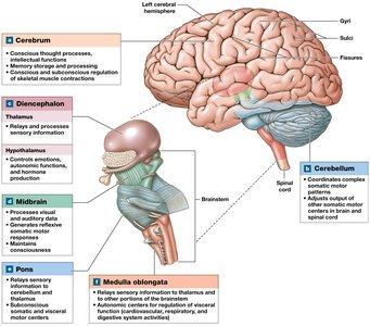
Brainstem
The brainstem connects the cerebrum and cerebellum to the spinal cord and is responsible for basic life functions. It includes the midbrain, pons, and medulla oblongata.
Midbrain: Processes visual and auditory information, maintains consciousness
Pons: Relays information to/from cerebellum, regulates breathing
Medulla oblongata: Regulates autonomic functions (heart rate, blood pressure)
Protection and Support of the Brain
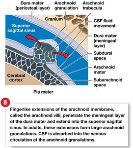
Cranial Meninges and Cerebrospinal Fluid (CSF)
The brain is protected by three layers of connective tissue called the cranial meninges (dura mater, arachnoid mater, pia mater) and by cerebrospinal fluid (CSF). The meninges stabilize and cushion the brain, while CSF provides buoyancy and protection against sudden movements.
Dural folds: Hold the brain in position
CSF: Cushions and supports the brain, circulates nutrients, removes waste
Clinical relevance: Cranial trauma, epidural and subdural hemorrhages can damage brain tissue

Blood Supply and Barriers
The brain receives nutrients and oxygen via the internal carotid and vertebral arteries. The blood-brain barrier (BBB) and blood-CSF barrier regulate the movement of substances between the blood and the brain, protecting neural tissue from toxins and pathogens.
BBB: Only lipid-soluble substances diffuse freely; glucose and ions are actively transported
Blood-CSF barrier: Formed by ependymal cells with tight junctions at the choroid plexus
Breaks in BBB: Occur in circumventricular organs for hormone release
Brainstem Components and Functions
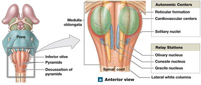
Medulla Oblongata
The medulla oblongata is the most inferior part of the brainstem, connecting the brain to the spinal cord. It contains nuclei for autonomic reflexes, cranial nerves, and relay stations for sensory and motor pathways.
Reflex centers: Cardiovascular and respiratory rhythmicity centers
Sensory/motor nuclei: Cranial nerves VIII, IX, X, XI, XII
Relay stations: Gracile and cuneate nuclei (somatic sensory), solitary nuclei (visceral sensory), inferior olivary complex (motor coordination)

Pons
The pons is located above the medulla and contains nuclei for cranial nerves V, VI, VII, and VIII. It regulates respiration and relays information between the cerebellum and other brain regions.
Respiratory centers: Apneustic and pneumotaxic centers
Tracts: Ascending, descending, and transverse pontine fibers

Midbrain
The midbrain (mesencephalon) processes visual and auditory information and maintains consciousness. It contains the tectum (corpora quadrigemina), tegmentum (red nucleus, substantia nigra), and cerebral peduncles.
Superior colliculi: Visual reflexes
Inferior colliculi: Auditory reflexes
Red nucleus: Motor coordination
Substantia nigra: Dopamine production, inhibits basal nuclei (affected in Parkinson's disease)

Cerebellum
Structure and Function
The cerebellum coordinates voluntary movements, balance, and posture. It contains the cerebellar cortex (gray matter), arbor vitae (white matter), and cerebellar peduncles (tracts connecting to other brain regions).
Lobes: Anterior, posterior, and flocculonodular
Vermis: Separates hemispheres
Disorders: Ataxia (loss of coordination), often due to trauma, stroke, or alcohol

Diencephalon
Thalamus
The thalamus is the main relay station for sensory information ascending to the cerebral cortex. It contains several nuclei with specialized functions, including relaying sensory and motor signals and integrating emotional and memory functions.
Anterior nuclei: Limbic system (emotions)
Medial nuclei: Emotional awareness
Ventral nuclei: Relay sensory and motor information
Lateral/medial geniculate bodies: Visual and auditory processing
Nuclei/Body | Function |
|---|---|
Anterior nuclei | Part of the limbic system |
Medial nuclei | Integrate sensory information for projection to the frontal lobes |
Ventral nuclei | Project sensory information to the primary sensory cortex; relay information from cerebellum and basal nuclei to motor area of cerebral cortex |
Lateral geniculate body | Projects visual information to the visual cortex; integrates sensory information and influences emotional states |
Medial geniculate body | Projects auditory information to the auditory cortex; integrates sensory information and influences emotional states |
Hypothalamus
The hypothalamus regulates homeostasis, emotions, and links the nervous and endocrine systems. It controls autonomic functions, hormone secretion, circadian rhythms, and behavioral drives such as hunger and thirst.
Hormones: ADH, oxytocin
Centers: Feeding, thirst, satiety, temperature regulation
Connections: Infundibulum (to pituitary), mammillary bodies (feeding reflexes)
Limbic System
Structure and Function
The limbic system is a functional grouping of structures involved in emotion, motivation, and memory. It links conscious and unconscious brain functions and facilitates memory storage and retrieval.
Components: Cingulate gyrus, hippocampus (learning, memory), amygdala (emotion, autonomic response), fornix (tract connecting hippocampus and hypothalamus)
Cerebrum: Structure and Functional Areas
Organization
The cerebrum is divided into lobes (frontal, parietal, temporal, occipital, insula) and contains both gray matter (cortex, basal nuclei) and white matter (fiber tracts). The cortex is responsible for processing sensory and motor information, as well as higher cognitive functions.
Gyri: Increase surface area for cortical neurons
Sulci: Separate lobes and regions
White matter tracts: Association, commissural, and projection fibers
Basal Nuclei
The basal nuclei are deep masses of gray matter involved in the subconscious regulation of movement and muscle tone. They coordinate learned movement patterns and provide feedback to the cortex.
Components: Caudate nucleus, putamen, globus pallidus
Functional Areas of the Cortex
Primary motor cortex: Controls voluntary skeletal muscles (precentral gyrus, frontal lobe)
Primary somatosensory cortex: Receives touch, pressure, pain, temperature (postcentral gyrus, parietal lobe)
Visual cortex: Occipital lobe
Auditory and olfactory cortex: Temporal lobe
Gustatory cortex: Insula
Association areas: Interpret sensory data, coordinate motor responses
Integrative centers: Prefrontal cortex (abstract thought), Wernicke's area (language comprehension), Broca's area (speech production)
Lobe/Area | Function |
|---|---|
Frontal lobe | Primary motor cortex: voluntary control of skeletal muscles |
Parietal lobe | Primary somatosensory cortex: conscious perception of touch, pressure, pain, vibration, taste, temperature |
Occipital lobe | Visual cortex: conscious perception of visual stimuli |
Temporal lobe | Auditory and olfactory cortex: conscious perception of hearing and smell |
All lobes | Integration and processing of sensory data; initiation of motor activities |
Hemispheric Lateralization
The left and right cerebral hemispheres have specialized functions. The left hemisphere is dominant for language, math, and logic, while the right hemisphere is involved in spatial analysis, recognition of faces, and emotional content of language.
Brain Waves and Electroencephalogram (EEG)
Brain activity can be measured by EEG, which records electrical patterns (brain waves):
Alpha waves: Awake, relaxed adults
Beta waves: Concentration, mental activity
Theta waves: Children, frustrated adults, or brain disorders
Delta waves: Deep sleep, brain damage in adults
Seizures are abnormal, synchronized electrical activity, and epilepsy is a disorder characterized by recurrent seizures.
Cranial Reflexes
Somatic and Visceral Reflexes
Cranial reflexes are automatic responses to stimuli involving cranial nerves. They can be monosynaptic or polysynaptic and are clinically useful for assessing brain and nerve function.
Reflex | Stimulus | Afferents | Central Synapse | Efferents | Response |
|---|---|---|---|---|---|
Corneal reflex | Contact with corneal surface | Trigeminal (V) | Motor nucleus for facial (VII) | Facial (VII) | Blinking of eyelids |
Tympanic reflex | Loud noise | Vestibulocochlear (VIII) | Inferior colliculus | Facial (VII) | Reduced movement of auditory ossicles |
Auditory reflexes | Loud noise | Vestibulocochlear (VIII) | Motor nuclei of brainstem and spinal cord | III, IV, VI, VII, X, cervical nerves | Eye/head movements to sound |
Vestibulo-ocular reflexes | Rotation of head | Vestibulocochlear (VIII) | Motor nuclei controlling eye muscles | III, IV, VI | Opposite movement of eyes to stabilize vision |
Pupillary reflex | Light in one eye | Optic (II) | Superior colliculus | Oculomotor (III) | Pupil constricts |
Consensual light reflex | Light in one eye | Optic (II) | Superior colliculus | Oculomotor (III) | Pupil constricts in both eyes |
